آخر المواضيع المضافة

 النبات
الحيوان
الأحياء المجهرية
علم الأمراض
التقانة الإحيائية
التقنية الحيوية المكروبية
التقنية الحياتية النانوية
علم الأجنة
الأحياء الجزيئي
علم وظائف الأعضاء
الغدد
المضادات الحيوية
النبات
الحيوان
الأحياء المجهرية
علم الأمراض
التقانة الإحيائية
التقنية الحيوية المكروبية
التقنية الحياتية النانوية
علم الأجنة
الأحياء الجزيئي
علم وظائف الأعضاء
الغدد
المضادات الحيوية| Light Microscopy |


|
|
Read More
Date: 21-10-2015
Date: 18-10-2015
Date: 21-10-2015
|
Light Microscopy
A light microscope (LM) is an instrument that uses visible light and magnifying lenses to examine small objects not visible to the naked eye, or in finer detail than the naked eye allows. Magnification, however, is not the most important issue in microscopy. Mere magnification without added detail is scientifically useless, just as endlessly enlarging a small photograph may not reveal any more detail, but only larger blurs. The usefulness of any microscope is that it produces better resolution than the eye. Resolution is the ability to distinguish two objects as separate entities, rather than seeing them blurred together as a single smudge. The history of microscopy has revolved largely around technological advances that have produced better resolution.
History of the Light Microscope
Light microscopes date at least to 1595, when Zacharias Jansen (1580-1638) of Holland invented a compound light microscope, one that used two lenses, with the second lens further magnifying the image produced by the first. His microscopes were collapsing tubes used like a telescope in reverse, and produced magnifications up to nine times (9x).
Antony van Leeuwenhoek (1632-1723) invented a simple (one-lens) microscope around 1670 that magnified up to 200 xs and achieved twice the resolution of the best compound microscopes of his day, mainly because he crafted better lenses. While others were making lenses by such methods as squashing molten glass between pieces of wood, Leeuwenhoek made them by carefully grinding and polishing solid glass. He thus became the first to see individual cells, including bacteria, protozoans, muscle cells, and sperm.
Englishman Robert Hooke (1635-1703) further refined the compound microscope, adding such features as a stage to hold the specimen, an illuminator, and coarse and fine focus controls. Until 1800, compound microscopes designed by Hooke and others were limited to magnifications of 30x to 50x, and their images exhibited blurry edges (spherical aberration) and rainbow-like distortions (chromatic aberration). The most significant improvement in microscope optics was achieved in the nineteenth century, when business partners Carl Zeiss (1816-1888) and Ernst Abbe (1840-1905) added the substage condenser and developed superior lenses that greatly reduced chromatic and spherical aberration, while permitting vastly improved resolution and higher magnification.
Tissue Preparation
The advancement of light microscopy also required methods for preserving plant and animal tissues and making their cellular details more visible, methods collectively called histotechnique (from histo, meaning “tissue”). In brief, classical histotechnique involves preserving a specimen in a fixative, such as formalin, to prevent decay; embedding it in a block of paraffin and slicing it very thinly with an instrument called a microtome; removing the paraffin with a solvent; and then staining the tissue, usually with two or more dyes. The slices of tissue, called histological sections, are typically thinner than a single cell. The colors of a prepared tissue are not natural colors, but they make the tissue’s structural details more visible. A widely used stain combination called hematoxylin and eosin, for example, typically colors cell nuclei violet and the cytoplasm pink.
Other methods of histotechnique have been developed for special purposes. One variation is to embed the tissue in special plastics (resins), allowing for thinner sectioning. Another is the frozen section method, in which a tissue is frozen with compressed carbon dioxide and sectioned with a special cold microtome, eliminating the time-consuming process of paraffin embedding. Some prefer this method for its relative simplicity, and its speed is an asset in hospitals, where a biopsied tissue may need to be examined rapidly and the diagnosis reported to the surgeon while the patient is in the operating room.
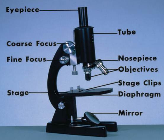
A compound light microscope.
Varieties of Light Microscopes
Most compound microscopes today have an illuminator built into the base. A condenser located below the stage has lenses that focus the light on the specimen and a diaphragm that regulates contrast. After passing through the specimen on the stage, the light enters an objective lens. Most light microscopes have three or four objective lenses on a rotating turret. These lenses magnify the image by 4x to 100x. The light then passes up the body tube to an ocular lens that magnifies the image another 10x to 15x. Research-grade microscopes and the better student microscopes have a pair of ocular lenses so that one can view the specimen with both eyes at once.
There are many varieties of compound light microscopes for special purposes. For viewing tissue cultures covered with liquid media, biologists can use an inverted light microscope in which the culture is illuminated from above and the objective lenses are positioned below the specimen. The phase contrast microscope can be used to enhance contrast in living specimens, thus avoiding the use of lethal fixatives and stains. The polarizing light microscope is used for analyzing crystals and minerals, among other things. The fluorescence microscope is used to examine structures that bind special fluorescent dyes. It can be used, for example, to identify where a dye- tagged hormone binds to its target cell.
Compound light microscopes achieve useful magnifications up to 1200x and resolutions down to about 0.25 micrometers. That is, two objects in a cell can be as close as 0.25 micrometers and still detected as separate entities. Such resolution is good enough to see most bacteria and some mitochondria and microvilli.
These microscopes generally require thin, transparent, relatively small specimens. They also require that the user adjust to the phenomenon of optical inversion; if a specimen is moved to the left, it appears under the microscope to move right; when moved up, it appears to move down; and vice versa. The stereomicroscope works at much lower magnification and resolution, but has several advantages: (1) it has two lens systems that view the specimen from slightly different angles, thus giving the specimen a stereoscopic (three-dimensional) appearance; (2) it can use either transmitted or reflected light; and with reflected light, it can be used to view opaque specimens such as rocks, fossils, insects, electronic circuit boards, and so forth; (3) it has a much greater working distance between the specimen and objective lens, allowing for the examination of relatively large objects and for easier manipulation of objects under the microscope; (4) the working distance enables relatively easy dissection of specimens such as insects, allowing hands and instruments to reach the working space while one looks through the microscope; and (5) it does not produce optical inversion; that is, movements to the right appear to go to the right, making dissection and other manipulations much easier.
The utility of light microscopy is governed by its use of visible light, which limits resolution. The shorter the wavelength of the illumination, the better the resolution. Electron beams have shorter wavelengths than photons. The invention of the electron microscope in the late 1930s and its refinement over the next half century permitted vastly improved visualization of cell and tissue fine structure.
References
Bradbury, Savile, and Brian Bracegirdle. Introduction to Light Microscopy. New York: Springer-Verlag, 1998.
Jones, Thomas E. History of the Light Microscope. <http://www.utmem.edu/~thjones/ hist/hist_mic.htm>.
Levine, S., and L. Johnstone. The Microscope Book. New York: Sterling Publishing Co., 1996.
Nachtigall, Werner. Exploring with the Microscope: A Book of Discovery and Learning. London: Sterling Publications, 1997.
Rogers, K. The Usborne Complete Book of the Microscope. Tulsa, OK: EDC Publishing, 1999.
WWW Virtual Library: Microscopy. <http://www.ou.edu/research/electron/mirror/>.


|
|
|
|
لشعر لامع وكثيف وصحي.. وصفة تكشف "سرا آسيويا" قديما
|
|
|
|
|
|
|
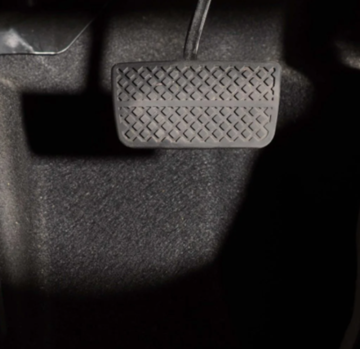
كيفية الحفاظ على فرامل السيارة لضمان الأمان المثالي
|
|
|
|
|
|
|
العتبة العباسية المقدسة تجري القرعة الخاصة بأداء مناسك الحج لمنتسبيها
|
|
|