Theory
Vibrational spectroscopy has been an integral tool for the identification and characterisation of drugs. When one thinks of vibrational spectroscopy, typically infrared (IR) techniques come to mind, not Raman spectroscopy. However, since the late 1980s a renaissance of the Raman technique has occurred, mainly through instrumentation development. These developments have led to unique applications in the pharmaceutical and forensic industries in which drug identification and characterisation are necessary. This chapter presents the theory, instrumentation, sampling techniques and applications of Raman spectroscopy as applied to drugs.
Raman spectroscopy is a form of vibrational spectroscopy that has widespread use in pharmaceutical investigations. Applications include chemical structure elucidation, routine chemical identification and solid-state characterisation, such as polymorphism. Raman spectroscopy is also applicable to drug product characterisation including solid form analysis of the drug incorporated into the formulation, contaminant analysis, drug–excipient interaction and problem solving. A distinct advantage of pharmaceutical analysis by Raman spectroscopy is the ease of the technique and its broad range of applicability. Analysis can be performed on virtually any type of sample, such as single crystals, bulk materials, slurries, creams, particulates, films, solutions (aqueous and organic), oils, gas-phase samples and on-process streams (the latter through the use of fibre-optic probes). Additionally, Raman spectroscopy is typically nondestructive in nature, and so the material can be recovered for further characterisation. Since Raman spectroscopy measures the vibrational motions associated with a molecule, it is complementary to IR spectroscopy as well as to other characterisation techniques. Finally, under proper sampling conditions, Raman spectroscopy is a quantitative technique.
When a compound is irradiated with mono chromatic radiation, the radiation is transmitted, absorbed or scattered by the molecule. As we have seen from the preceding chapters, UV and IR spectroscopy are primarily concerned with absorbed and transmitted radiation. However, it is the scattered radiation that is of interest in Raman spectroscopy. Of the scattered radiation, a majority of the photons are scattered at the same frequency as the incident radiation frequency. This form of scattering has been termed elastic or Rayleigh scattering. If the scattered radiation is passed into a spectrometer, a strong Rayleigh line is detected at the unmodified frequency of radiation used to excite the sample. Additionally, a very small proportion of photons (about one per million) are scattered at frequencies arrayed above and below the frequency of the Rayleigh line.
The differences between the incident frequency of radiation and arrayed frequencies correspond to the frequency of molecular vibrations present in the molecules of the sample. These wavelength-shifted frequencies are due to what is termed inelastic scattering, and a collection of this wavelength shifted frequencies is termed a Raman spectrum. The frequencies of molecular vibrations are typically 1012 to 1014 Hz. A more convenient unit, which is proportional to frequency, is wavenumber (cm-1) since fundamental vibrational modes lie between 3600 and 50 cm-1. For example, a Raman line at 2980 cm-1 may be obtained on either side of the Rayleigh line and thus the sample possesses a vibrational mode at this frequency.
As shown in Figure 1, when a molecule is irradiated with monochromatic radiation, a number of different transitions may occur. If the radiation is of sufficient energy, an absorption process may occur that represents an electronic transition (S0 →S1, UV-visible spectroscopy). If a slightly less energetic source is used, the molecule is promoted to a virtual state. The virtual state represents a distortion of the electron distribution of a covalent bond within the molecule. After promotion to the virtual state, the molecule immediately relaxes back to the original ground electronic state by emitting a photon. If the molecule relaxes back to the original vibrational state, the emitted photon is of the same frequency as the incident radiation. This represents Rayleigh or elastic scattering. If the molecule relaxes back to a higher vibrational energy state, the emitted photon represents less energy than the incident radiation. The inelastically scattered photon has a longer wavelength (lower frequency) than the incident radiation. This energy transition represents Stokes Raman scattering. Conversely, if the molecule relaxes back to a lower vibrational energy state, the emitted photon represents greater energy than the incident radiation. The inelastically scattered photon has a shorter wavelength (higher frequency) than the incident radiation. This energy transition represents anti-Stokes Raman scattering. Generally, the anti-Stokes lines are less intense than the Stokes lines because these transitions arise from higher vibrational energy levels that will be populated in only a small proportion of molecules, as described by the Boltzmann distribution. Hence, the Stokes portion of the spectrum is generally used.
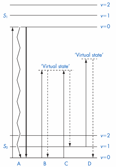
Figure 1 Jablonski energy level diagram illustrating possible transitions. Solid lines represent absorption processes and dotted lines represent scattering processes. A, electronic transition with nonradiative decay (heat, zig zag line) or radiative decay (fluorescence, thick line); B, Rayleigh scattering; C, Stokes Raman transition; D, anti-Stokes Raman transition. S0 is the singlet ground state, S1 the lowest singlet excited state and v represents vibrational energy levels within each electronic state.
A Raman spectrum is normally represented as a plot of Raman scattering intensity (ordinate, y axis) versus wavelength (abscissa, x-axis). Normally, the abscissa of the spectrum is labelled as wavenumber shift or Raman shift (cm1) and the negative sign (for Stokes shift) is omitted (Fig. 2). The wavenumber or Raman shift represents the shift in frequency of a photon from the exciting wavelength. Pharmaceutically and forensically relevant molecules are typically covalently bound organic molecules. The chemical bonds within these molecules comprise an electron cloud. In a Raman experiment, the electromagnetic radiation incident on the molecule consists of oscillating electric and magnetic fields. Interaction of the electromagnetic radiation with the chemical bond causes the electron cloud to oscillate. The oscillation, in turn, causes a photon to be emitted, which is called scattering. In Rayleigh scattering, the energy from the incident electromagnetic radiation causes electron oscillation and the emitted photon is observed at the same frequency. In Raman scattering, an additional energy transition occurs. The polarisability of the electron cloud may change as the position of the atoms that make up the chemical bond changes. In other words, a vibrational mode may cause atoms to be displaced, which in turn affects the polarisability of the electron cloud of the chemical bond. In this case, the incident radiation causes the electron cloud to oscillate, but the electron cloud oscillation is also affected by the change in polarisability caused by a change in the position of the atoms during the molecular vibration. Oscillation causes a photon to be emitted, but the frequency is perturbed by the change in polarisability of the chemical bond. For a vibrational mode to be Raman active, a change in polarisability must take place during the vibration. This is termed the Raman selection rule. Since the polarisability of a chemical bond is dependent upon the atoms making up that bond, as well as atoms in close proximity, Raman spectroscopy is a probe into the chemical or physical structure of a molecule. This is why Raman spectroscopy is an important tool for pharmaceutical analysis.
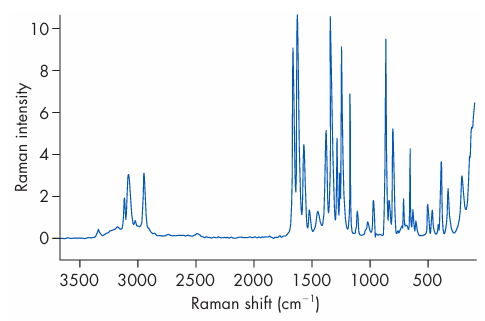
Figure 2 FT-Raman spectrum of paracetamol (acetaminophen).








