آخر المواضيع المضافة

 النبات
الحيوان
الأحياء المجهرية
علم الأمراض
التقانة الإحيائية
التقنية الحيوية المكروبية
التقنية الحياتية النانوية
علم الأجنة
الأحياء الجزيئي
علم وظائف الأعضاء
الغدد
المضادات الحيوية
النبات
الحيوان
الأحياء المجهرية
علم الأمراض
التقانة الإحيائية
التقنية الحيوية المكروبية
التقنية الحياتية النانوية
علم الأجنة
الأحياء الجزيئي
علم وظائف الأعضاء
الغدد
المضادات الحيوية| Cytosol |


|
|
Read More
Date: 2-11-2016
Date: 28-10-2015
Date: 15-11-2016
|
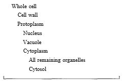
Cytosol
Most of the volume of cytoplasm is a clear substance called cytosol or hyaloplasm. It is mostly water, enzymes, and the numerous chemical precursors, intermediates, and products of enzymatic reactions (Table 1). Within cytosol are free ribosomes (not attached to HER), as well as skeletal structures the microtubules and microfilaments.
TABLE 1: Subunits of the Cell
MICROTUBULES
Microtubules (Fig. 1) are the most abundant and easily studied of the structural elements of a cell, and they have many functions. They act as a "cytoskeleton," holding certain regions of the cell surface back while other parts expand. Without them, cells would be just spheres, but by reinforcing specific areas, cell growth and expansion are directed to weaker areas. In other cases, microtubules assemble into arrays like an antenna which either catch vesicles and guide them to specific sites or cover a region, thereby excluding the vesicles (Fig. 2).
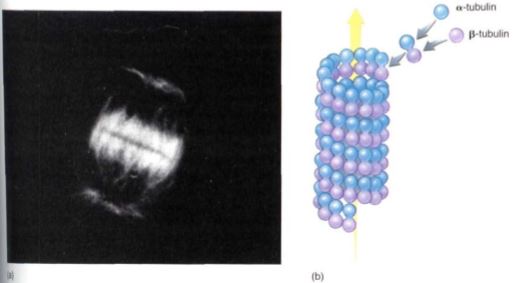
FIGURE 1: (a) Microtubules that are part of a dividing nucleus and are involved in pulling chromosomes to the ends of the cells. A fluorescent dye was used to stain only microtubules, so no other organelles are visible (X 7000). (Courtesy of Dr. Kevin C. Vaughn, Southern Weed Science Laboratory) (b) Alpha and beta tubulin associate into a dimer called tubulin, and dimers aggregate into a microtubule. When no longer needed, microtubules depolymerize back to the monomers, which are recycled to build other microtubules.
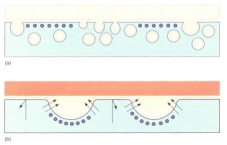
FIGURE 2: Microtubules (depicted here in purple) are often located next to the plasma membrane. (a) They may act as a screen that keeps vesicles away from the plasma membrane, or (b) they may pull the plasma membrane away from the cell wall, allowing material to accumulate there.
Finally, microtubules are the means of motility for both organelles and whole cells. The framework that moves chromosomes during division of the nucleus is composed of microtubules, and microtubules can attach to and move whole nuclei, mitochondria, and other organelles .
Microtubules are composed of just two types of globular protein, alpha tubulin and beta tubulin, which associate as dimers called tubulin that further crystallize into a straight tubule with a diameter of 20 to 25 nm. This is a reversible process; when a microtubule is no longer needed, it depolymerizes back into its component monomers, which disperse into the cytosol until the cell needs to assemble a new microtubule. Control of polymerization and depolymerization is not well-understood. When microtubules occur as individuals or small clusters, new tubulin dimers are added to or removed from one end automatically. In other instances, microtubules occur in large clusters, often in a highly ordered arrangement, and a small body is usually associated with the orderly production of microtubules. For example, when the nucleus undergoes division, an array of microtubules called the spindle is formed in the middle of the cell, and spindle microtubules push and pull the chromosomes to their proper positions.
In all animals and in some fungi and algae, a pair of organelles called centrioles is associated with the formation of the spindle. A centriole is made up of nine sets of three short microtubules (Fig. 3); the nine triplets are held together by fine protein spokes.
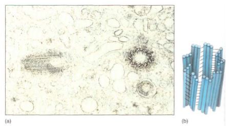
FIGURE 3: (a) The two members of a pair of centrioles are always located perpendicular to each other. Here, one is seen in transverse section, the other in longitudinal section (X 100,000). (Biology Media) (b) Each part of a centriole contains a circle of nine sets of three microtubules attached to each other by fine filaments along their sides
Centrioles were assumed to be responsible for the organization and polymerization of the spindle microtubules even though plants never have centrioles. At present, however, centrioles are suspected to have nothing to do with spindle formation, even in animals. Rather, it may be that the pair of centrioles may be separated by the growth of the spindle, which pushes them to opposite ends of the cell so that each daughter cell receives one during cell division.
Much more elaborate sets of precisely arranged microtubules occur in cilia (sing.: cilium) and flagella (sing.: flagellum) (Figs. 4 and 5), which appear to be identical except that cilia are short (about 2 pm) and occur in groups, whereas flagella tend to be much longer (up to several micrometers) and usually occur either singly or in sets of two or four. Both are present on many types of algae and motile fungi, but in plants only the sperm cells have flagella. In flowering plants and conifers, no cells, not even sperm cells, ever have cilia or flagella.
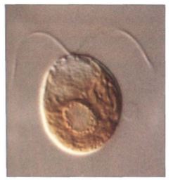
FIGURE 4: Flagella occur on many algal and fungal cells, especially the unicellular organisms. This is the alga Dunalliela. Most cells of multicellular organisms are not flagellated, but their sperm cells are (X500).
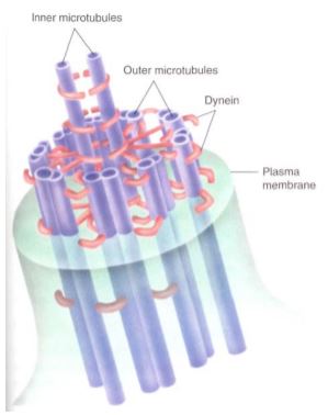
FIGURE 5: Cilia and flagella are composed of two central single microtubules surrounded by nine sets of two microtubules. The doublets are not merely attached to each other; they actually share tubulin monomers. The outer member of each doublet has two short arms.
In cross-section each cilium or flagellum has a "9 + 2" arrangement: Nine pairs of fused microtubules surround two individual microtubules. The outer doublets each have two arms composed of the protein dynein, and each doublet is connected to the central pair of microtubules by protein spokes. The dynein arms convert the chemical energy of ATP into kinetic energy and bend, "walking" along the adjacent microtubule doublet. One set of doublets slides relative to the adjacent set, causing that side of the cilium or flagellum to become shorter and bend the structure. Then, as another set of microtubules slides, the cilium or flagellum bends in a different direction. The result is a powerful beating motion. If the cilia or flagella are on small organisms such as algae, fungi, or protozoans, the organism swims rapidly and gracefully.
Cilia and flagella can polymerize and depolymerize rapidly, but they never form autonomously; each is always associated with a basal body. Basal bodies appear to be identical to centrioles by electron microscopy. It was assumed that basal bodies organize the formation of flagellar microtubules, but recent studies have shown that as a flagellum grows, new monomers of tubulin and dynein are added to the tip, not the base where the basal body is located. The exact relationship between flagella and basal bodies is still not known.
MICROFILAMENTS
Like microtubules, microfilaments are constructed by the assembly of globular proteins -in this case just one type, actin. Microfilaments are narrower than microtubules (only 3 to 6 nm in diameter), and they have been implicated tentatively in different types of structure and movement.


|
|
|
|
اكتشاف تأثير صحي مزدوج لتلوث الهواء على البالغين في منتصف العمر
|
|
|
|
|
|
|
زهور برية شائعة لتر ميم الأعصاب التالفة
|
|
|
|
|
|
جمعيّة العميد وقسم الشؤون الفكريّة تدعوان الباحثين للمشاركة في الملتقى العلمي الوطني الأوّل
|
|
|
|
الأمين العام المساعد لجامعة الدول العربية السابق: جناح جمعية العميد في معرض تونس ثمين بإصداراته
|
|
|
|
المجمع العلمي يستأنف فعاليات محفل منابر النور في واسط
|
|
|
|
برعاية العتبة العباسيّة المقدّسة فرقة العبّاس (عليه السلام) تُقيم معرضًا يوثّق انتصاراتها في قرية البشير بمحافظة كركوك
|