آخر المواضيع المضافة

 النبات
الحيوان
الأحياء المجهرية
علم الأمراض
التقانة الإحيائية
التقنية الحيوية المكروبية
التقنية الحياتية النانوية
علم الأجنة
الأحياء الجزيئي
علم وظائف الأعضاء
الغدد
المضادات الحيوية
النبات
الحيوان
الأحياء المجهرية
علم الأمراض
التقانة الإحيائية
التقنية الحيوية المكروبية
التقنية الحياتية النانوية
علم الأجنة
الأحياء الجزيئي
علم وظائف الأعضاء
الغدد
المضادات الحيوية| DNA Glycosylases |


|
|
Read More
Date: 17-5-2016
Date: 23-12-2015
Date: 6-4-2021
|
DNA Glycosylases
Base excision repair is the system for repairing abnormal bases in DNA. The abnormal bases and simple base lesions are removed from DNA by the combined actions of DNA glycosylases (1-3) and AP (apurinic/apyrimidinic) endonucleases. The basic reaction carried out by glycosylases is the cleavage of the glycosylic bond joining the base to deoxyribose. Some of the glycosylases cleave both the glycosylic bond and the phosphodiester bond 3′ to the resulting AP site in a more or less concerted reaction. Hence, these DNA glycosylases have been classified as either “pure” glycosylases or as glycosylase/AP lyases.
1. Uracil DNA Glycosylases
Uracil in DNA arises from two sources: (i) direct misincorporation into DNA in place of thymine by DNA polymerases and (ii) deamination of cytosine. Most DNA polymerases cannot discriminate between dTTP and dUTP; hence the rates of incorporation of thymine and uracil into DNA are proportional to the concentrations of the corresponding dNTPs in the nucleotide pool. The dTTP:dUTP ratio is about 1000:1, so about one uracil is incorporated into each Okazaki fragment during DNA replication. The second source of uracil in DNA is that produced by deamination of cytosine. Cytosine deaminates at normal pH and temperature at a rate of 10–16s–1 for double-stranded DNA and at a rate of 10–10s–1 for single-stranded DNA. The deamination rate is greatly accelerated by low pH, high temperature, and oxidants. Uracil in DNA is removed by uracil glycosylases. Two have been identified in Escherichia coli and in humans (4): (i) uracil glycosylase and (ii) the double-strand-specific thymine glycosylase, which has also been shown to act on uracil residues in double-stranded DNA.
1.1. Uracil DNA Glycosylase (UDG(
The classic UDG is a 25- to 30-kDa monomeric protein with no cofactor or metal ion requirement. The enzyme is highly conserved between prokaryotes and eukaryotes, and it is the major uracil glycosylase in most organisms examined. The enzyme acts on single- and double-stranded DNA with equal efficiency. UDG is one of the several known DNA modification/repair enzymes that during catalysis flip out the base from inside the helix into a cavity within the enzyme (5-7). After cleaving off the base, the enzyme dissociates from DNA. This UDG has no detectable AP lyase activity.
1.2. Thymine–DNA Glycosylase (TDG(
In humans, this enzyme specifically removes thymines from T-G mismatches and uracils from U-G mismatches within double-stranded DNA (4-6). It is thought that the major source of mismatched thymines in mammalian cells is deamination of 5-methylcytosine bases, which creates a T-G mismatch. Because about 20% of cytosine bases are methylated in mammalian cells, even low-frequency deamination of MeCyt generates high levels of T-G mismatches. Hence it is likely that TDG plays an important role in preventing mutations and maintaining genomic stability. Even though DNA glycosylases in general, and UDG in particular, are considered to be highly specific enzymes, recent work has shown that UDG removes from double-stranded DNA many oxidized pyrimidines generated by ionizing radiation, and even normal pyrimidines (7). The Km and kcat catalytic parameters for removal of these lesions were found to be within the physiologically relevant range, so it is possible that UDG and TDG also perform auxiliary functions in repairing DNA damage caused by oxidative stress, in addition to their primary roles.
2. Thymine Glycol DNA Glycosylase
The prototype of thymine glycol DNA glycosylases is E. coli endonuclease III (8). It is a 40-kDa polypeptide chain with a [4Fe–4S] cluster (9) (see Iron–Sulfur Proteins). This enzyme has been detected in various organisms by one of its multiple activities and, accordingly, was given various names: endonuclease III, UV endonuclease, redoxendonuclease, and others. The enzyme has been highly conserved during evolution. Enzymes with sequence and functional homology to E. coli endonuclease III exist in yeast and humans (10). These enzymes have a relatively wide substrate spectrum and act on ring-saturated, ring-contracted, and ring-rearranged pyrimidines. The crystal structure of E. coli endonuclease III has been determined (11). The enzyme flips out of damaged base into the active site and cleaves the glycosylic bond and the 3′ phosphodiester bond of the resulting AP site, before dissociating from DNA. The iron–sulfur center within the protein has no catalytic role. Catalysis does not involve redox chemistry, but it occurs by a simple lyase reaction involving the -amino group of a lysine residue.
3. Methylpurine DNA Glycosylase (MPG)
The MPG enzyme was originally identified as “3-methyladenine DNA glycosylase”; hence it is also referred to by that name (12, 13). There are two enzymes in E. coli with MPG activity, one that has a narrow substrate spectrum called TagI, and a second, TagII, with a much greater substrate range (14) . The eukaryotic enzymes, including that in humans, are more similar to TagII with respect to their substrate preference than to TagI of E. coli. MPGs act on 3-methyladenine, 7-methyladenine, and O4-methylthymine. They clearly also repair alkylated bases other than methyl purines, and hence “alkylated purine DNA glycosylase” would perhaps be a more appropriate name for this group of enzymes. They have no overt AP lyase activity.
4. 8-Oxoguanine Glycosylase
This enzyme was first identified as “formamidinopyrimidine DNA glycosylase” for the name of the substrate (FAPy) that is generated from guanine by ionizing radiation (15). Subsequently, it was found that the enzyme is very active on 8-oxoguanine, which is generated in vast quantities in the DNA by ionizing radiation and by oxidative stress. The enzyme is a 20-kDa monomer with no requirement for cofactors or divalent cations. The enzyme is widespread in the biological world, having been found in E. coli, yeast, and humans. It is a glycosylase and an AP lyase. In addition to cleaving the 3′ phosphodiester bond by b-elimination, it also cleaves the 5′ phosphodiester bond by d-elimination. Thus, this is a unique glycosylase that performs glycosylase/b-d-elimination by a concerted mechanism; in doing so, it generates a one-nucleotide gap.
5. Pyrimidine Dimer Glycosylase
This enzyme has been found in two sources thus far: T4 phage and Micrococcus luteus. The enzymes from the two sources share sequence homology and are presumed to act by the same mechanism. Both are 18-kDa monomers, with no cofactor and no requirement for divalent cations.
The enzyme cleaves the glycosylic bond of the 5′ base of cyclobutane dimer and the intradimer phosphodiester bond by b-elimination. Thus, the cleavage generates a 3′-OH end and a 5′ terminus with a dangling pyrimidine dimer (16). The 5′ end must be further processed by a 5′- to 3′-exonuclease before DNA polymerase can produce a ligatable product. The structure of T4 endonuclease V complexed with substrate indicates that the enzyme flips out one of the adenine bases opposite the pyrimidine dimer (17). This binding mechanism explains the high specificity for pyrimidine dimers in double-stranded DNA.
6. A-G Mismatch DNA Glycosylase
The 8-oxoguanine lesion is a frequent product of ionizing radiation and oxidative damage to DNA. Most often, 8-oxoguanine mispairs with adenine during replication. The resulting adenine-8-oxoguanine mispair is the substrate for A-G mismatch DNA glycosylase (18). This 30-kDa enzyme has limited sequence similarity to endonuclease III (thymine glycol endonuclease) and also has a [4Fe–4S] center. The protein in E. coli is encoded by the mutY gene; hence the enzyme is also referred to as MutY glycosylase. The enzyme also cleaves A residues in A-G mismatches, albeit at a lower efficiency than A-8-oxoG mismatches. In addition to glycosylase activity, it also possesses AP lyase activity and thus cleaves the 3′ glycosylic bond 3′ to an AP site, either as part of glycosylase/AP lyase concerted reaction or as an independent AP lyase acting on an isolated AP site.
References
1. T. Lindahl (1974) Proc. Natl. Acad. Sci. USA 71, 3649–3653.
2. T. Lindahl (1976) Nature 259, 64–66.
3. J. Laval (1977) Nature 269, 828–832.
4. P. Gallinari and J. Jiricny (1996) Nature 383, 735–738.
5. S. Klimasauskas, S. Kumar, R. J. Roberts, and X. Cheng (1994) Cell 76, 357–369.
6. T. E. Barrett, R. Savva, G. Panayotou, T. Barlow, T. Brown, J. Jiricny, and L. H. Pearl (1998( Cell 92, 117–129.
7. K. G. Berdal, R. F. Johansen, and E. Seeberg (1998) EMBO J. 17, 363–367.
8. B. Demple and S. Linn (1980) Nature 287, 203–208.
9. R. P. Cunningham, H. Asahara, J. F. Bank, C. P. Scholes, J. C. Salerno, K. Surerus, E. Munck, J. McCracken, J. Peisach, and M. H. Emptage (1988) Biochemistry 28, 4450–4455.
10. T. P. Hilbert, W. Chaung, R. J. Boorstein, R. P. Cunningham, and G. W. Teebor (1997) J. Biol. Chem. 272, 6733–6740.
11. C. Kuo, D. E. McRee, C. L. Fisher, S. F. O''Handley, R. P. Cunningham, and J. A. Tainer (1992) Science 258, 434–440.
12. Y. Nakabeppu, H. Kondo, and M. Sekiguchi (1984) J. Biol. Chem. 259, 13723–13729.
13. G. Evensen and E. Seeberg (1982) Nature 296, 773–775.
14. J. Labahn, O. D. Scharer, A. Long, K. Ezaz-Nikapy, G. L. Verdine, and T. E. Ellenberger (1996) Cell 86, 321–329.
15. S. Boiteux, T. R. O''Connor, and J. Laval (1987) EMBO J. 6, 3177–3183.
16. W. A. Haseltine, L. K. Gordon, C. P. Lindan, R. H. Grafstrom, N. L. Shaper, and L. Grossman (1980) Nature 285, 634–641.
17. D. G. Vassylyev, T. Kashiwagi, Y. Mikami, M. Ariyoshi, S. Iwai, E. Ohtsuka, and K. Morikawa (1995) Cell 83, 773–782.
18. K. G. Au, S. Clark, J. H. Miller, and P. Modrich (1989) Proc. Natl. Acad. Sci. USA 86, 8871–8881.


|
|
|
|
اكتشاف تأثير صحي مزدوج لتلوث الهواء على البالغين في منتصف العمر
|
|
|
|
|
|
|
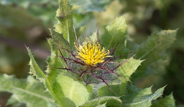
زهور برية شائعة لتر ميم الأعصاب التالفة
|
|
|
|
|
|
جمعيّة العميد وقسم الشؤون الفكريّة تدعوان الباحثين للمشاركة في الملتقى العلمي الوطني الأوّل
|
|
|
|
الأمين العام المساعد لجامعة الدول العربية السابق: جناح جمعية العميد في معرض تونس ثمين بإصداراته
|
|
|
|
المجمع العلمي يستأنف فعاليات محفل منابر النور في واسط
|
|
|
|
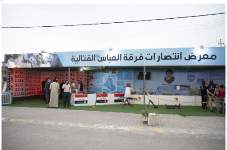
برعاية العتبة العباسيّة المقدّسة فرقة العبّاس (عليه السلام) تُقيم معرضًا يوثّق انتصاراتها في قرية البشير بمحافظة كركوك
|