
آخر المواضيع المضافة

 النبات
الحيوان
الأحياء المجهرية
علم الأمراض
التقانة الإحيائية
التقنية الحيوية المكروبية
التقنية الحياتية النانوية
علم الأجنة
الأحياء الجزيئي
علم وظائف الأعضاء
الغدد
المضادات الحيوية
النبات
الحيوان
الأحياء المجهرية
علم الأمراض
التقانة الإحيائية
التقنية الحيوية المكروبية
التقنية الحياتية النانوية
علم الأجنة
الأحياء الجزيئي
علم وظائف الأعضاء
الغدد
المضادات الحيوية| Chlamydia |


|
|
Read More
Date: 18-3-2016
Date: 14-3-2016
Date: 3-3-2016
|
Chlamydia
Chlamydiae are obligate cell parasites. They go through two stages in their reproductive cycle: the elementary bodies (EB) are optimized to survive outside of host cells. In the form of the initial bodies (IB), the chlamydiae reproduce inside the host cells. The three human pathogen species of chlamydiae are C. psittaci, C. trachomatis, and C. pneumoniae. Tetracyclines and macrolides are suitable for treatment of all chlamydial infections.
C. psittaci is the cause of psittacosis or ornithosis. This zoonosis is a systemic disease of birds. The pathogens enter human lungs when dust containing chlamydiae is inhaled. After an incubation period of one to three weeks, pneumonia develops that often shows an atypical clinical course.
C. trachomatis is found only in humans. This species causes the following diseases: 1. Trachoma, a chronic follicular kerato-conjunctivitis. The pathogens are transmitted by smear infection. 2. Inclusion conjunctivitis in newborn children and swimming-pool conjunctivitis. 3. Nonspecific urogenital infections in both men and women (urethritis, cervicitis, salpingitis, etc.). 4. Lymphogranuloma venereum, a venereal disease observed mainly in countries with warm climates.
C. pneumoniae is responsible for infections of the upper respiratory tract as well as for a mild form of pneumonia. There is current discussion in the literature concerning a possible role of C. pneumoniae in the pathogenesis of atherosclerotic cardiovascular disease.
Overview and General Characteristics of Chlamydiae
Definition and classification. The bacteria in the taxonomic family Chlamy- diaceae are small (0.3-1 µm) obligate cell parasites with a Gram-negative cell wall. The reproductive cycle of the chlamydiae comprises two developmental stages: The elementary bodies are optimally adapted to survival outside of host cells. The initial bodies, also known as reticulate bodies, are the form in which the chlamydiae reproduce inside the host cells by means of transverse fission. Three human pathogen species of chlamydiae are known:
C. psittaci, C. trachomatis (with the biovars trachoma and lymphogranuloma venereum), and C. pneumoniae.
Morphology and developmental cycle. Two morphologically and functionally distinct forms are known:
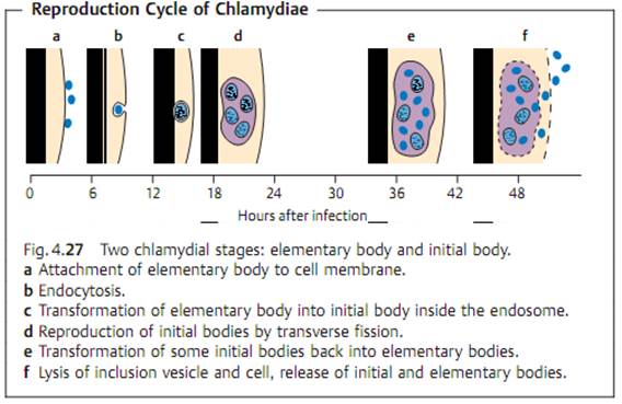
- Elementary bodies. The round to oval, optically dense elementary bodies have a diameter of approximately 300 nm. They represent the infectious form of the pathogen and are specialized for the demands of existence outside the host cells. Once the elementary bodies have attached themselves to specific host cell receptors, they invade the cells by means of endocytosis (Fig. 4.27). Inside the cell, they are enclosed in an endocytotic membrane vesicle or inclusion, in which they transform themselves into the other form-initial bodies-within a matter of hours.
- Initial bodies. Chlamydiae in this spherical to oval form are also known as reticular bodies. They have a diameter of approximately 1000 nm. The initial bodies reproduce by means of transverse fission and are not infectious while in this stage. At the end of the cycle, the initial bodies are transformed back into elementary bodies. The cell breaks open and releases the elementary bodies to continue the cycle by attaching themselves to new host cells.
Culture. Chlamydiae exploit energy metabolism processes in their host cells that they themselves are lacking (ATP synthesis). For this reason, they can only be grown in special cell cultures, in the yolk sacs of embryonated hen eggs, or in experimental animals.
Chlamydia psittaci (Ornithosis, Psittacosis)
Pathogenesis and clinical picture. The natural hosts of C. psittaci are birds. This species causes infections of the respiratory organs, the intestinal tract, the genital tract, and the conjunctiva of parrots and other birds. Humans are infected by inhalation of dust (from bird excrements) containing the pathogens, more rarely by inhalation of infectious aerosols. After an incubation period of one to three weeks, ornithosis presents with fever, headache, and a pneumonia that often takes an atypical clinical course. The infection may, however, also show no more than the symptoms of a common cold, or even remain clinically silent. Infected persons are not usually sources of infection.
Diagnosis. The pathogen can be grown from sputum in special cell cultures. Direct detection in the culture is difficult and only possible in specially equipped laboratories. The complement binding reaction can be used to identify antibodies to a generic antigen common to all chlamydiae, so that this test would also have a positive result in the presence of other chlamydial infections. The antibody test of choice is indirect microimmunofluorescence.
Therapy. Tetracyclines (doxycycline) and macrolides.
Epidemiology and prevention. Ornithosis affects birds worldwide. It is also observed in poultry. Diagnosis of an ornithosis in a human patient necessitates a search for and elimination of the source, especially if the birds in question are household pets.
Chlamydia trachomatis (Trachoma, Lymphogranuloma venereum)
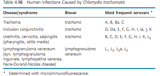
C. trachomatis is a pathogen that infects only humans. Table 4.16 lists the relevant diseases, biovars, and serovars.
Trachoma is a follicular keratoconjunctivitis. The disease occurs in all climatic zones, although it is more frequent in warmer, less-developed countries. It is estimated that 400 million people carry this chronic infection and that it has caused blindness in six million. The pathogen is transmitted by direct contact and indirectly via objects in daily use. Left untreated, the initially acute inflammation can develop a chronic course lasting months or years and leading to formation of a corneal scar, which can then cause blindness. The laboratory diagnostics procedure involves detection of C. trachomatis in conjuncival smears using direct immunofluorescence microscopy. The fluoro-chrome-marked monoclonal antibodies are directed against the MOMP (major outer membrane protein) of C. trachomatis. The pathogen can also be grown in cell cultures. The therapeutic method of choice is systemic and local application of tetracycline over a period of several weeks.
Inclusion conjunctivitis. This is an acute, purulent papillary conjunctivitis that may affect neonates, children, and adults (swimming-pool conjunctivitis). Newborn children are infected during birth by pathogens colonizing the birth canal. Left untreated, a pannus may form as in trachoma, followed by corneal scarring. Laboratory diagnosis and therapy as in trachoma.
Genital infections. C. trachomatis is responsible for 30-60% of cases of non-gonococcal urethritis (NGU) in men. Possible complications include prostatitis and epididymitis. The pathogens are communicated by venereal transmission.The source of infection is the female sexual partner, who often shows no clinical symptoms.
In women, C. trachomatis can cause urethritis, proctitis, or infections of the genital organs. It has even been known to cause pelvioperitonitis and perihepatitis. Massive perinatal infection of a neonate may lead to an interstitial chlamydial pneumonia.
The relevant diagnostic tools include:
1. Detection under the microscope in smear material using direct immuno-fluorescence (see under trachoma).
2. Direct identification by means of amplification of a specific DNA sequence in smear material and urine.
3. Growing in special cell cultures.
Lymphogranuloma venereum. This venereal disease (syn. lymphogranuloma inguinale, lymphopathia venerea (Favre-Durand-Nicolas disease) not to be confused with granuloma inguinale) is frequently observed in the inhabitants of warm climatic zones. A herpetiform primary lesion develops at the site of invasion in the genital area, which then becomes an ulcus with accompanying lymphadenitis. Laboratory diagnosis is based on isolating the proliferating pathogen in cell cultures from purulent material obtained from the ulcus or from matted lymph nodes. The antibodies can be identified using the complement binding reaction or the microimmunofluorescence test. tetracycline and macrolides are the potentially useful antibiotic types.
Chlamydia pneumoniae
This new chlamydial species (formerly TWAR chlamydiae) causes infections of the respiratory organs in humans that usually run a mild course: influenza like infections, sinusitis, pharyngitis, bronchitis, pneumonias (atypical). Clinically silent infections are frequent. C. pneumoniae is pathogenic in humans only. The pathogen is transmitted by aerosol droplets. These infections are probably among the most frequent human chlamydial infections. Serological studies have demonstrated antibodies to C. pneumoniae in 60% of adults. Specific laboratory diagnosis is difficult. Special laboratories can grow and identify the pathogen in cultures and detect it under the microscope using marked antibodies to the LPS (although this test is positive for all chlamydial infections). C. pneumoniae-specific antibodies can be identified with the microimmunofluorescence method. In a primary infection, a measurable titer does not develop for some weeks and is also quite low. The antibiotics of choice are tetracyclines or macrolides. There is a growing body of evidence supporting a causal contribution by C. pneumoniae to atherosclerotic plaque in the coronary arteries, and thus to the pathogenesis of coronary heart disease.


|
|
|
|
إجراء أول اختبار لدواء "ثوري" يتصدى لعدة أنواع من السرطان
|
|
|
|
|
|
|
دراسة تكشف "سببا غريبا" يعيق نمو الطيور
|
|
|
|
|
|
لأعضاء مدوّنة الكفيل السيد الصافي يؤكّد على تفعيل القصة والرواية المجسّدة للمبادئ الإسلامية والموجدة لحلول المشاكل المجتمعية
|
|
|
|
قسم الشؤون الفكرية يناقش سبل تعزيز التعاون المشترك مع المؤسّسات الأكاديمية في نيجيريا
|
|
|
|
ضمن برنامج عُرفاء المنصّة قسم التطوير يقيم ورشة في (فنّ الٕالقاء) لمنتسبي العتبة العباسية
|
|
|
|
وفد نيجيري يُشيد بمشروع المجمع العلمي لحفظ القرآن الكريم
|