
آخر المواضيع المضافة

 النبات
الحيوان
الأحياء المجهرية
علم الأمراض
التقانة الإحيائية
التقنية الحيوية المكروبية
التقنية الحياتية النانوية
علم الأجنة
الأحياء الجزيئي
علم وظائف الأعضاء
الغدد
المضادات الحيوية
النبات
الحيوان
الأحياء المجهرية
علم الأمراض
التقانة الإحيائية
التقنية الحيوية المكروبية
التقنية الحياتية النانوية
علم الأجنة
الأحياء الجزيئي
علم وظائف الأعضاء
الغدد
المضادات الحيوية| Heterochromatin Depends on Interactions with Histones |


|
|
Read More
Date: 17-5-2016
Date: 9-5-2021
Date: 8-12-2015
|
Heterochromatin Depends on Interactions with Histones
KEY CONCEPTS
- HP1 is the key protein in forming mammalian heterochromatin; it acts by binding to methylated histone H3 and leads to the formation of higher-order chromatin structures.
- Rap1 initiates formation of heterochromatin in yeast by binding to specific target sequences in DNA.
- The targets of Rap1 include telomeric repeats and silencers at HML and HMR.
- Rap1 recruits Sir3 and Sir4, which interact with the Nterminal tails of H3 and H4.
- Sir2 deacetylates the N-terminal tails of H3 and H4 and promotes spreading of Sir3 and Sir4.
- RNAi pathways promote heterochromatin formation at centromeres.
Inactivation of chromatin occurs via a combination of covalent modifications and the addition of proteins to the nucleosomal fiber. The inactivation may be due to a variety of effects, including condensation of chromatin to make it inaccessible to the apparatus needed for gene expression, addition of proteins that directly block access to regulatory sites, or proteins that directly inhibit transcription.
Two systems that have been characterized at the molecular level involve HP1 in mammals and the SIR complex in yeast. Although many of the proteins involved in each system are not evolutionarily related, the general reaction mechanism is similar: The points of contact in chromatin are the N-terminal tails of the histones.
Insight into the molecular mechanisms that regulate the formation of heterochromatin originated with mutants that affect PEV. Twenty-eight genes have been identified in Drosophila that affect PEV. They are named systematically as Su(var) for genes whosepr oducts act to suppress variegation and E(var) for genes whose products enhance variegation. These genes were named for the behavior of the mutant loci; thus, Su(var) mutations lie in genes whose products are needed for the formation of heterochromatin. They include enzymes that act on chromatin, such as histone deacetylases, and proteins that are localized to heterochromatin. In contrast, E(var) mutations lie in genes whose products are needed to activate gene expression. They include members of the SWI/SNF chromatin remodeling complex .
HP1 (heterochromatin protein 1) is one of the most important Su(var) proteins. It was originally identified as a protein that is localized to heterochromatin by staining polytene chromosomes with an antibody directed against the protein. It was later shown to be the product of the gene Su(var)2–5. Its homolog in the yeast Schizosaccharomyces pombe is encoded by swi6. HP1 is now called HP1α because two related proteins, HP1β and HP1γ, have
since been found.
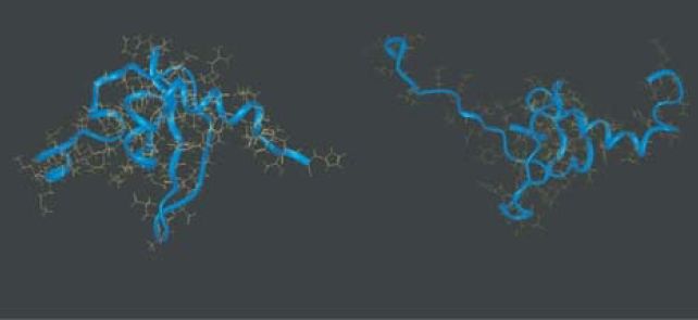
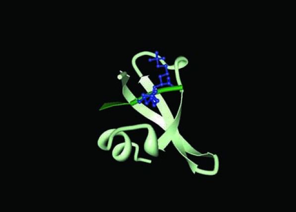
HP1 contains a chromodomain near the N-terminus and another domain that is related to it (the chromo shadow domain) at the Cterminus. HP1 is able to interact with many chromosomal proteins through the chromo shadow domain while the HP1 chromodomain binds to histone H3 that is dimethylated or trimethylated at lysine 9 (H3K9me3). FIGURE 1 shows the structures of the chromodomain and chromo shadow domains of HP1, as well as a structure showing the interaction between the chromodomain and the methylated lysine. This interaction is a hallmark of inactive
chromatin.

(a)
(b)
(c)
FIGURE 1 (a, b) HP1 contains a chromodomain and a chromo shadow domain. (c)Trimethylation of histone H3 K9 creates a binding site for HP1.
(a, b) Photo reproduced from G. Lomberk, L. Wallrath, and R. Urrutia, Genome Biol. 7 (2006): p. 228. Used with permission of Raul A. Urrutia and Gwen Lamberk, Mayo Clinic. (c) Structure from Protein Data Bank 1KNE. S. A. Jacobs and S. Khorasanizadeh, Science 275 (2002): 2080–2083.
Mutation of a deacetylase that acts on H3K14Ac prevents the methylation at K9, resulting in loss of the HP1 binding site. This suggests the model for initiating formation of heterochromatin shown in FIGURE 2. First the deacetylase acts to remove the modification at K14, and this allows the SUV39H1 methyltransferase (also known as KMT1A) to methylate H3K9 to create the methylated signal to which HP1 will bind. FIGURE 3 shows that the inactive region may then be extended by the ability of further HP1 molecules to interact with one another.
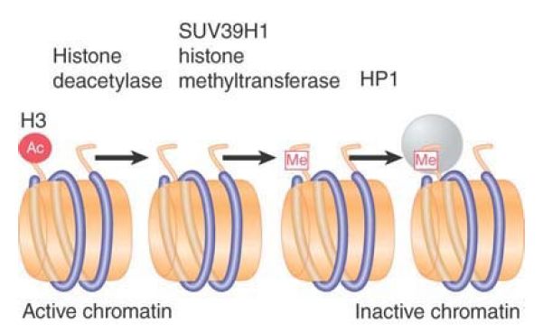
FIGURE 3. SUV39H1 is a histone methyltransferase that acts on K9 of histone H3. HP1 binds to the methylated histone.
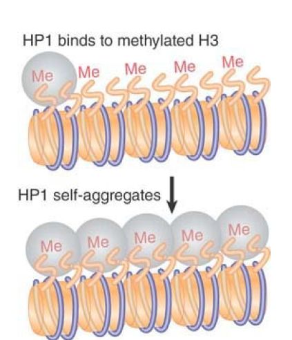
FIGURE 3. Binding of HP1 to methylated histone H3 forms a trigger for silencing because additional molecules of HP1 aggregate along the methylated chromatin domain.
The state of histone methylation is important in the control of heterochromatin or euchromatin states. Methylation of H3K9 demarcates heterochromatin, whereas H3K4 methylation demarcates euchromatin. A trimethyl H3K4 demethylase found in S.pombe referred to as Lid2 interacts with the Clr4 H3K9 methyltransferase, resulting in H3K4 hypomethylation and heterochromatin formation. The link between H3K4 demethylation and H3K9 methylation suggests that the two reactions act in a coordinated manner to control the relative state of heterochromatin or euchromatin of a specific region.
Heterochromatin formation at telomeres and silent mating-type loci in yeast relies on an overlapping set of genes known as silent information regulators (SIR genes). Binding of SIR proteins can actually silence any promoter or coding region, but under normal conditions nucleation or the recruitment of SIR proteins to specific sequences allows for silencing to be targeted to specific regions of the genome—specifically the telomeres and HM loci. Mutations in SIR2, SIR3, or SIR4 cause HML and HMR to become activated and also relieve the inactivation of genes that have been integrated near telomeric heterochromatin. The products of these SIR genes therefore function to maintain the inactive state of both types of heterochromatin.
FIGURE 4 shows a model for the actions of these proteins. Only one of them—Rap1—is a sequence-specific DNA-binding protein. It binds to the C A repeats at the telomeres and also binds to the cis-acting silencer elements that are needed for repression of HML and HMR. The proteins Sir3 and Sir4 interact with Rap1 and also with one another (they may function as a heteromultimer). Sir3 and Sir4 interact with the N-terminal tails of the histones H3 and H4, with a preference for unacetylated tails. Another SIR protein, Sir2, is a deacetylase, and its activity is 1–3 necessary to maintain binding of the Sir3/Sir4 complex to chromatin.
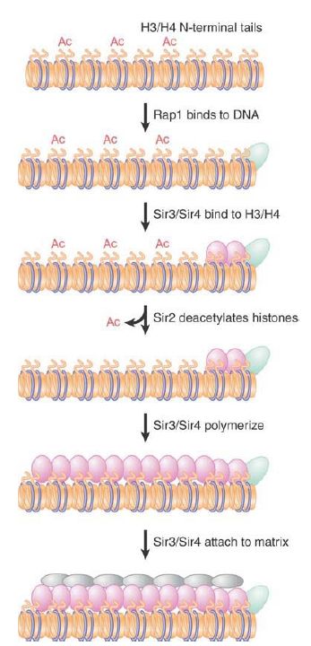
FIGURE 4. Formation of heterochromatin is initiated when Rap1 binds to DNA. Sir3/4 bind to Rap1 and also to histones H3/H4. Sir2 deacetylates histones. The SIR complex polymerizes along chromatin and may connect telomeres to the nuclear matrix.
Rap1 has the crucial role of identifying the DNA sequences at which heterochromatin forms. It recruits Sir4, which, in turn, recruits both its binding partner Sir3 and the HDAC Sir2. Sir3 and Sir4 then interact directly with histones H3 and H4. Once Sir3 and Sir4 have bound to histones H3 and H4, the complex (including Sir2) can polymerize further and spread along the chromatin fiber. This may inactivate the region, either because coating with the Sir3/Sir4 complex itself has an inhibitory effect, or because Sir2-dependent deacetylation represses transcription. It is not known what limits the spreading of the complex. The C-terminus of Sir3 has a similarity to nuclear lamin proteins (constituents of the nuclear matrix) and may be responsible for tethering heterochromatin to the nuclear periphery.
A similar series of events forms the silenced regions at HMR and HML. Three sequence-specific factors are involved in triggering formation of the complex: Rap1, Abf1 (a transcription factor), and the origin replication complex (ORC). In this case, Sir1 (which is not required for telomeric silencing) binds to a sequence-specific factor and recruits Sir2, -3, and -4 to form the repressive structure. As at the telomeres, Sir2-dependent deacetylation is necessary to maintain binding of the SIR complex to chromatin.
Formation of heterochromatin in the yeast S. pombe utilizes an RNAi-dependent pathway (see the Regulatory RNA chapter). This pathway is initiated by the production of siRNA molecules resulting from transcription of centromeric repeats. These siRNAs result in formation of the RNA-induced transcriptional silencing (RITS) complex. The siRNA components are responsible for localizing the complex at centromeres. The complex contains proteins that are homologs of those involved in heterochromatin formation in other organisms, including plants, Caenorhabditis elegans, and D. melanogaster. This complex includes Argonaute, which is involved in targeting RNA-induced silencing complex (RISC) remodeling complexes to chromatin. The siRNA complex promotes methylation of histone H3K9 by the Clr4 methyltransferase (also known as KMT1, a homolog of Drosophila Su[Var]3–9). H3K9 methylation recruits the S. pombe homolog of HP1, Swi6.
How does a silencing complex repress chromatin activity? It could condense chromatin so that regulator proteins cannot find their targets. The simplest case would be to suppose that the presence of a silencing complex is mutually incompatible with the presence of transcription factors and RNA polymerase. The cause could be that silencing complexes block remodeling (and thus indirectly prevent factors from binding) or that they directly obscure the binding sites on DNA for the transcription factors. The situation may not be that simple, though, because transcription factors and RNA polymerase can be found at promoters in silenced chromatin. This could mean that the silencing complex prevents the factors from working rather than from binding as such. In fact, competition may exist between gene activators and the repressing effects of chromatin so that activation of a promoter inhibits spread of the silencing complex.
Centromeric heterochromatin is particularly interesting, because it is not necessarily nucleated by simple sequences (as is the case for telomeres and the mating-type loci in yeast), but instead
depends on more complex mechanisms, some of which are RNAi dependent. The specialized chromatin structure that forms at the centromere may be associated with the formation of heterochromatin in the region. The unique centromeric chromatin structure and the centromere-specific histone H3 variants are discussed in the Chromosomes and Chromatin chapters. In human cells, the centromere-specific protein CENP-B is required to initiate modifications of histone H3 (deacetylation of K9 and K14, followed by methylation of K9) that trigger an association with HP1 that leads to the formation of heterochromatin in the region. Moreover, heterochromatin and RNAi are required to establish the human CenH3 homolog, CENP-A, at centromeres. Heterochromatin is often present near CENP-A chromatin and the RNAi-directed
heterochromatin flanking the central kinetochore domain is required for kinetochore assembly. Several factors, such as the Suv39 methyltransferase, HP1, and components of the RNAi pathway .
Studies of the propagation of the pathogenic yeast Candida albicans have shown that naked centromeric DNA that can confer centromeric activity in vivo is not able to assemble functional centromeric chromatin de novo when reintroduced into cells. This suggests that C. albicans centromeres are dependent on their preexisting chromatin state and provides an example of epigenetic propagation of a centromere.


|
|
|
|
إجراء أول اختبار لدواء "ثوري" يتصدى لعدة أنواع من السرطان
|
|
|
|
|
|
|
دراسة تكشف "سببا غريبا" يعيق نمو الطيور
|
|
|
|
|
|
لأعضاء مدوّنة الكفيل السيد الصافي يؤكّد على تفعيل القصة والرواية المجسّدة للمبادئ الإسلامية والموجدة لحلول المشاكل المجتمعية
|
|
|
|
قسم الشؤون الفكرية يناقش سبل تعزيز التعاون المشترك مع المؤسّسات الأكاديمية في نيجيريا
|
|
|
|
ضمن برنامج عُرفاء المنصّة قسم التطوير يقيم ورشة في (فنّ الٕالقاء) لمنتسبي العتبة العباسية
|
|
|
|
وفد نيجيري يُشيد بمشروع المجمع العلمي لحفظ القرآن الكريم
|