
oxicological screening , Quantification of carboxyhaemoglobin in blood by spectrophotometry
 المؤلف:
Sue Jickells , Adam Negrusz (Editors)
 المصدر:
Clarkes Analytical Forensic Toxicology
 الجزء والصفحة:
234
 2026-10-18
 81
oxicological screening
Quantification of carboxyhaemoglobin in blood by spectrophotometry
Principle When a reducing agent (sodium dithionite) is added to the blood, both the oxygenated form and the methaemoglobin are converted quantitatively to the reduced form, which has the visible spectrum B shown in Fig. 1. Carbon monoxide has a much greater affinity for haemoglobin than does oxygen, and the COHb is not reduced by sodium dithionite. Thus, even when treated with sodium dithionite, COHb retains its normal twin-peaked spectrum, marked A in Fig. 8.1. The maximal difference between the spectra of A and B is at 540 nm, while at 579 nm the spectra have the same absorbance (isosbestic point). The percentage saturation of carbon monoxide in a blood sample (A) can be calculated from measurements of the absorbance of the carbon monoxide-free sample (B) and the untreated sample (C), after reduction of each with sodium dithionite. Standards Gas bottles of pure carbon monoxide can be obtained. Alternatively, commercial reference standards of haemolysed blood in sealed glass ampoules are available (IL, Warrington, UK).
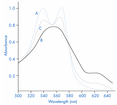
Figure 1 Ultraviolet spectra of (A) carboxyhaemo globin, (B) reduced haemoglobin and (C) a blood sample from a patient poisoned with carbon monoxide.
 0
 0
 الاكثر قراءة في تحليل السموم
 اخر الاخبار
اخبار العتبة العباسية المقدسة








