


النبات

مواضيع عامة في علم النبات
الجذور - السيقان - الأوراق
النباتات الوعائية واللاوعائية
البذور (مغطاة البذور - عاريات البذور)
الطحالب
النباتات الطبية
الحيوان
مواضيع عامة في علم الحيوان
علم التشريح
التنوع الإحيائي
البايلوجيا الخلوية
الأحياء المجهرية
البكتيريا
الفطريات
الطفيليات
الفايروسات
علم الأمراض
الاورام
الامراض الوراثية
الامراض المناعية
الامراض المدارية
اضطرابات الدورة الدموية
مواضيع عامة في علم الامراض
الحشرات
التقانة الإحيائية
مواضيع عامة في التقانة الإحيائية
التقنية الحيوية المكروبية
التقنية الحيوية والميكروبات
الفعاليات الحيوية
وراثة الاحياء المجهرية
تصنيف الاحياء المجهرية
الاحياء المجهرية في الطبيعة
أيض الاجهاد
التقنية الحيوية والبيئة
التقنية الحيوية والطب
التقنية الحيوية والزراعة
التقنية الحيوية والصناعة
التقنية الحيوية والطاقة
البحار والطحالب الصغيرة
عزل البروتين
هندسة الجينات
التقنية الحياتية النانوية
مفاهيم التقنية الحيوية النانوية
التراكيب النانوية والمجاهر المستخدمة في رؤيتها
تصنيع وتخليق المواد النانوية
تطبيقات التقنية النانوية والحيوية النانوية
الرقائق والمتحسسات الحيوية
المصفوفات المجهرية وحاسوب الدنا
اللقاحات
البيئة والتلوث
علم الأجنة
اعضاء التكاثر وتشكل الاعراس
الاخصاب
التشطر
العصيبة وتشكل الجسيدات
تشكل اللواحق الجنينية
تكون المعيدة وظهور الطبقات الجنينية
مقدمة لعلم الاجنة
الأحياء الجزيئي
مواضيع عامة في الاحياء الجزيئي
علم وظائف الأعضاء
الغدد
مواضيع عامة في الغدد
الغدد الصم و هرموناتها
الجسم تحت السريري
الغدة النخامية
الغدة الكظرية
الغدة التناسلية
الغدة الدرقية والجار الدرقية
الغدة البنكرياسية
الغدة الصنوبرية
مواضيع عامة في علم وظائف الاعضاء
الخلية الحيوانية
الجهاز العصبي
أعضاء الحس
الجهاز العضلي
السوائل الجسمية
الجهاز الدوري والليمف
الجهاز التنفسي
الجهاز الهضمي
الجهاز البولي
المضادات الميكروبية
مواضيع عامة في المضادات الميكروبية
مضادات البكتيريا
مضادات الفطريات
مضادات الطفيليات
مضادات الفايروسات
علم الخلية
الوراثة
الأحياء العامة
المناعة
التحليلات المرضية
الكيمياء الحيوية
مواضيع متنوعة أخرى
الانزيمات

Fibroblasts-Fibrocytes
 المؤلف:
Kuehnel, W
المؤلف:
Kuehnel, W
 المصدر:
Color Atlas of Cytology, Histology, and Microscopic Anatomy
المصدر:
Color Atlas of Cytology, Histology, and Microscopic Anatomy
 الجزء والصفحة:
 10-1-2017
 3638
3638


+
-
20
Fibroblasts-Fibrocytes
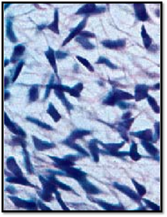
Fibrocytes are local (fixed) connective tissue cells. They are branched and connect to each other via cytoplasmic processes of different sizes. Other wise, the appearances of f ibrocytes differ, dependent on the type of the connective tissue and their function. In the usual sections, they attach so tightly to the surrounding connective tissue fibers that it of ten renders their cytoplasm invisible. The name fibroblast shows that the connective tissue cell has a specific functional role. Fibroblasts play an important role in the synthesis of extracellular substances ( extracellular matrix ), as in fibrillogenesis. This figure shows strongly basophilic f ibroblasts in the connective tissue of a fetal jawbone.
Stain: hemalum-eosin; magnification: × 500
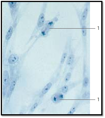
Fibroblasts from the edge fog of a cell culture ( cover-glass culture ). After seeding a cell culture, the propagating cells will spread. Their spreading in a sparse, thin layer to the underside of the cover g lass allows a microscopic examination. Fibroblasts are stretched, flattened cells. Their cytoplasmic processes may look like a membrane, or form spikes. They feature large, usually oval nuclei with prominent nucleoli and display a very delicate chromatin structure. The cytoplasm appears vitreous and is only lightly stained. Occasionally, it contains small fat droplets and vacuoles. Note the cells in mitosis 1 .
Stain: methylene blue; magnification: × 400
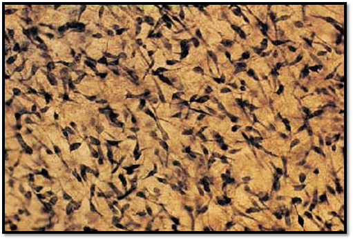
Fibrocytes from the connective tissue of a human amnion. Some of the oval or spindle-shaped fibrocytes have long processes, which will make contact with processes from other f ibroblasts. Whole-mount preparation. Stain: Heidenhain iron hematoxylin; magnification: × 50
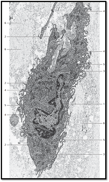
Fibrocyte from the epineurium of the median nerve with arcuate, slender processes 5 of different lengths. Fibrocytes tend to have the shape of a spindle and consequently, their nuclei are elongate d and of ten lobed 1 . The electron-dense, finely granulate d cytoplasm contains many small mitochondria 2 with an electron-dense, osmiophilic matrix and close to the surface, vacuoles of various sizes 4 . The granular endoplasmic reticulum 3 and the Golgi apparatus are only poorly developed. Close to the f ibrocytes—i.e., in the extracellular matrix of the connective tissue—there are multitudes of collagen fibrils 6 , which are all cut vertical to their axis. Fibroblasts and f ibrocytes are always present in connective tissue. They are important for the formation of fibers and the synthesis of nonstructural intercellular substances ( glycosaminoglycans). The cells discharge procollagen molecules into the extracellular space, which assemble to tropocollagen and finally to microfibrils. Different stimuli convert f ibrocytes back to f ibroblasts. They will then synthesize structural and nonstructural extracellular substances, for example during wound healing.
1 Lobed nucleus
2 Mitochondria
3 Granular ER
4 Vesicles
5 Cell processes in the form of tentacles and microvilli
6 Basic substance (extracellular matrix) with collagen fibrils
Electron microscopy; magnification: × 7000
References
Kuehnel, W.(2003). Color Atlas of Cytology, Histology, and Microscopic Anatomy. 4th edition . Institute of Anatomy Universitätzu Luebeck Luebeck, Germany . Thieme Stuttgart · New York .
 الاكثر قراءة في علم الخلية
 اخر الاخبار
اخبار العتبة العباسية المقدسة

الآخبار الصحية


مواضيع ذات صلة
 قسم الشؤون الفكرية يصدر كتاباً يوثق تاريخ السدانة في العتبة العباسية المقدسة
 "المهمة".. إصدار قصصي يوثّق القصص الفائزة في مسابقة فتوى الدفاع المقدسة للقصة القصيرة
 (نوافذ).. إصدار أدبي يوثق القصص الفائزة في مسابقة الإمام العسكري (عليه السلام)


