آخر المواضيع المضافة

 النبات
الحيوان
الأحياء المجهرية
علم الأمراض
التقانة الإحيائية
التقنية الحيوية المكروبية
التقنية الحياتية النانوية
علم الأجنة
الأحياء الجزيئي
علم وظائف الأعضاء
الغدد
المضادات الحيوية
النبات
الحيوان
الأحياء المجهرية
علم الأمراض
التقانة الإحيائية
التقنية الحيوية المكروبية
التقنية الحياتية النانوية
علم الأجنة
الأحياء الجزيئي
علم وظائف الأعضاء
الغدد
المضادات الحيوية| Cerebellar Cortex |


|
|
Read More
Date: 12-1-2017
Date: 18-1-2017
Date: 4-8-2016
|
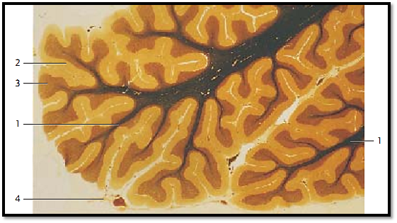
Cerebellar Cortex
Central sagittal section of the cerebellar vermis.
The cerebellar cortex is the about 1-mm thick folded layer of gray matter overlying the cerebellar surface. Grooves subdivide the gray matter in many small coils. The medullary layer of white matter consists of thin medullary layers 1 . In a medullar y sheath preparation, the cerebellar cortex shows an outer molecular layer (stratum moleculare) (stained yellow) 2 and an inner granular layer (stratum granulosum) (stained brownish) 3 . The layer between the inner and outer layers is the wide stratum ganglion are (stratum neuronorum piriformium gangliosum) 3 . This layer is easily recognized. It consists of large Purkinje cells (Purkinje cell layer). Remnants of the soft piamater 4 are visible on the surface of this section.
1 Medullar y layer
2 Molecular layer
3 Stratum ganglionare, Purkinje cell layer
4 Piamater
Stain: medullary sheath stain (modified Weiger t hematoxylin stain); magnification: × 5
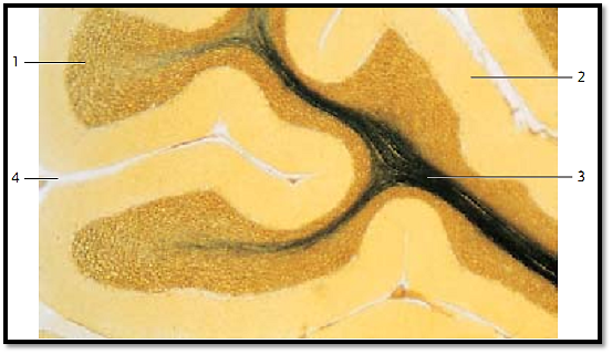
Cerebellar Cortex
Staining clearly differentiates between the granular layer 1 and the molecular layer 2 , which contains few cells and appears pale. The molecular layer is 430 μm thick , the granular layer measures about 350 μm at the turn of the coils. The layer is clearly thinner in the grooves. Numerous densely packed cells form the granular layer 1 . The granular structure is base d on the crowded presence of small cells. In general staining procedures, only the cell nuclei are clearly rendered. The Purkinje cells of the stratumganglionare ( stratum neuronorum piriformium) are located between the granular and molecular layers. The medullary layer 3 contains the myelinated nerve fibers of the cerebellar tracts.
1 Granular layer
2 Molecular layer
3 Medullar y layer with laminae
4 Piamater
Stain: Weiger t medullar y sheath staining; magnification: × 10
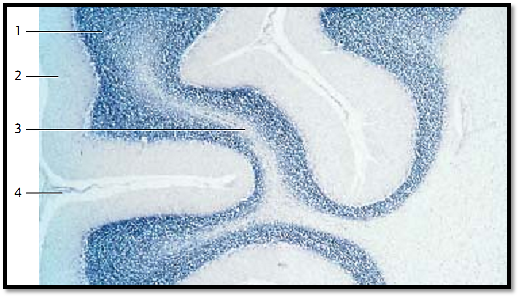
Cerebellar Cortex
Detail from a center sagittal section of the cerebellar vermis . The figure shows the result of Nissl staining of the nerve and glial cells ( Nissl image ). The cell-rich granular layer 1 is rendered particularly well, while the molecular layer is only slightly tinge d with a grayish blue hue. The laminae of the medullary layer 3 have remained unstained.
1 Granular layer
2 Molecular layer
3 Medullar y layer, laminae
4 Piamater
Stain: Nissl stain; magnification: × 10
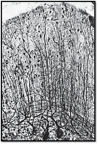
Cerebellar Cortex
The perikarya of the Purkinje cells in the stratum ganglionare are about 30 μm thick . They are arranged in a row. Purkinje cells appear striking even with general staining methods because they are by far the largest neurons of the cerebellum.
The round or rounded Purkinje cell bodies are darkly stained in this preparation. From their somata ascends an elaborately branched dendritic tree into the molecular layer and the glial external limiting membrane. The usually strong primary dendrites branch into secondary and tertiary dendrites. They form an espalier-like configuration in the molecular layer. The territories of individual Purkinje cells may overlap. There are fiber tracts in the lower third of the molecular layer immediately over the cell body of the Purkinje cells, which run parallel to the granular layer and vertical to the dendritic trees of the Purkinje cells. These fiber tracts are axons of basket cells, which are of ten called tangential f ibers. There are many small astrocytes and basket cells in the molecular layer (stained black). The lower edge of the figure shows the granular layer , which is tightly packed with small granular cells. The axons of the Purkinje cells (not sectioned) end as sole efferent fibers of the cerebellar cortex adjacent to the neurons of the cerebellar nuclei.
Stain: Bielschowsky -Gros silver impregnation; magnification: × 200
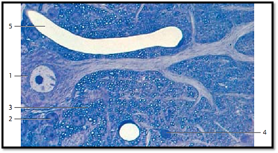
Cerebellar Cortex
Purkinje cells from the cerebellar cortex of a rhesus monkey, vermis, lobulus II. The primary dendrite of a Purkinje cell 1 ascends to the molecular layer and forms secondary and tertiary dendritic branches in this layer. The nuclei of granular cells 2 from the granular layer are locate d adjacent to the body of the Purkinje cell. A layer of cross-sectioned myelinated nerve fibers 3 in the lower third of the molecular layer follows. These are myelinated parallel f ibers , which are abundant in some parts of the cerebellum. Only the very large myelinated fibers in the plexus supraganglionare represent retrogressive collaterals of the Purkinje cell axons. At the magnification, the very fine parallel fibers are not visible in this figure in the upper two thirds of the molecular layer. There is a cross-sectioned capillary at the lower edge of the figure. The oligodendrocyte 4 next to it was identified by its dense cytoplasm.
1 Cell body of a Purkinje cell
2 Nucleus of a granular cell
3 Myelinated axons
4 Oligodendrocyte
5 Capillary
Semi-thin section; stain: methylene blue-azure II; magnification: × 630
References
Kuehnel, W.(2003). Color Atlas of Cytology, Histology, and Microscopic Anatomy. 4th edition . Institute of Anatomy Universitätzu Luebeck Luebeck, Germany . Thieme Stuttgar t · New York .


|
|
|
|
إجراء أول اختبار لدواء "ثوري" يتصدى لعدة أنواع من السرطان
|
|
|
|
|
|
|
دراسة تكشف "سببا غريبا" يعيق نمو الطيور
|
|
|
|
|
|
لأعضاء مدوّنة الكفيل السيد الصافي يؤكّد على تفعيل القصة والرواية المجسّدة للمبادئ الإسلامية والموجدة لحلول المشاكل المجتمعية
|
|
|
|
قسم الشؤون الفكرية يناقش سبل تعزيز التعاون المشترك مع المؤسّسات الأكاديمية في نيجيريا
|
|
|
|
ضمن برنامج عُرفاء المنصّة قسم التطوير يقيم ورشة في (فنّ الٕالقاء) لمنتسبي العتبة العباسية
|
|
|
|
وفد نيجيري يُشيد بمشروع المجمع العلمي لحفظ القرآن الكريم
|