
آخر المواضيع المضافة

 النبات
الحيوان
الأحياء المجهرية
علم الأمراض
التقانة الإحيائية
التقنية الحيوية المكروبية
التقنية الحياتية النانوية
علم الأجنة
الأحياء الجزيئي
علم وظائف الأعضاء
الغدد
المضادات الحيوية
النبات
الحيوان
الأحياء المجهرية
علم الأمراض
التقانة الإحيائية
التقنية الحيوية المكروبية
التقنية الحياتية النانوية
علم الأجنة
الأحياء الجزيئي
علم وظائف الأعضاء
الغدد
المضادات الحيوية| Golgi Apparatus |


|
|
Read More
Date: 2-8-2016
Date: 4-1-2017
Date: 1-8-2016
|
Golgi Apparatus
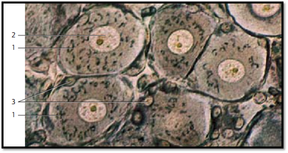
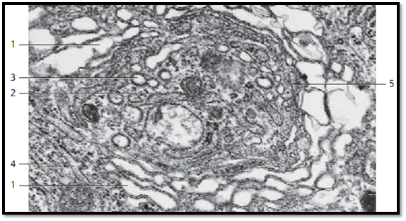
The Golgi apparatus was name d after Camillo Golgi (Nob el Prize 190 6), who discovered this cell structure in nerve cells (189 8) and assigned it the role of a cell organelle. The Golgi apparatus is ubiquitous in cells. The figure shows spinal ganglion cells with cytoplasmic structures in the form of black rods, hooks or loops 1 . In differentiate d polar cells from exocrine glands, they are locate d in the apical third of the cell, frequently in close proximity to secretory granules. Inspire d by its appearance, Golgi named this cell structure the inner reticular apparatus (“apparato reticolare in-terno”). The Golgi apparatus is not stained in routine histological preparations. However, components of the Golgi apparatus can re duce metal ions.
1 Golgi apparatus (apparato reticolare interno)
2 Ganglia nucleus with clearly visible nucleolus
3 Nuclei in satellite cells
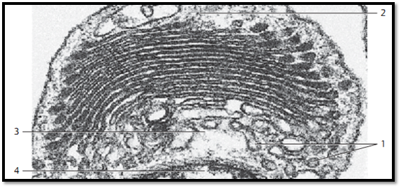
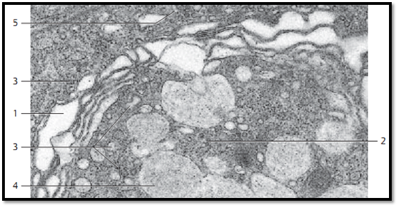
The Golgi apparatus consists of membranes that are about 6–8 nm thick . The basic unit of the Golgi apparatus is the dictyosome or Golgi field . In this sec-tion, it consists of a stack of 3–8 smooth (i.e., ribosome-free) slightly arcuate stacked membranes in close proximity to each other. The membranes en-close long narrow cisternae, which are slightly wider at both ends. The dictyosome can be compare d to a stack of flat membrane sacs with osmiophilic content ( export proteins ). Golgi cisternae are always accompanied by Golgi vesicles 1 , which deliver and export material (transport vesicles). The Golgi apparatus has therefore two faces, a convex (cis-), or forming face 2 and a concave trans-face ,or secretory face 3 . Prespermatid from Eisenia foetida.
1 Golgi vesicle (transpor t vesicle)
2 Convex, cis-face
3 Concave, trans -face
4 Nucleus, partial section
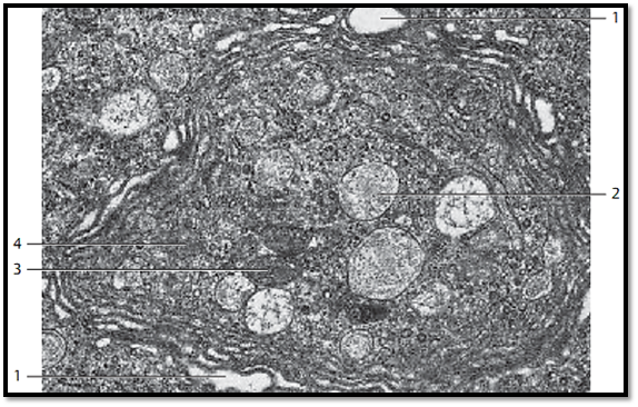
Large Golgi apparatus with smooth double membranes and Golgi vacuoles . Some of the cisternae are distended 1 . At the concave side (trans-face ) of the cisternae, there are very small vesicles (transport vesicles); among them are coated vesicles, but also somewhat larger vacuoles. They contain variable amounts of secretor y products. The more or less sharply curve d membrane stacks of the Golgi complex feature a convex cis-face ( uptake side) and a concave trans-face ( export side). These two distinct sides also contain different sets of enzymes.
1 Distended Golgi cisternae
2 Secretor y granules
3 Mitochondria
4 Golgi vesicles (transport vesicles)
The Golgi apparatus of ten contains several dictyosomes (Golgi fields). In cells with a polar structure, dictyosomes are usually localize d in the supranuclear region. The basic structural unit of the Golgi apparatus, however, is the Golgi cisterna. It is a flat, 1–2 μm wide membrane sac, which is often dilate d and fenestrate d at the outer edges. Several of these cisternae form a functional dictyosome. All dictyosomes in a cell combine d make up the Golgi apparatus. The ultrastructure of the Golgi apparatus ( Golgi complex) is distinct in its de-tail. The electron-microscopic examination of thin sections of cells reveals the Golgi apparatus as a characteristically structured system of smooth double membranes ( sacculi ), which are either arrange d in parallel stacks, or are layered around vacuoles. The membranes do not communicate with each other. Of ten, 6–8 membrane profiles form a stack of flat or arcuate cisternae. Most of them are distended at the ends, like flasks. At those ends are small vesicles (transport vesicles) and vacuoles, which have been pinched off from the distended cisternae ( sacculi) .
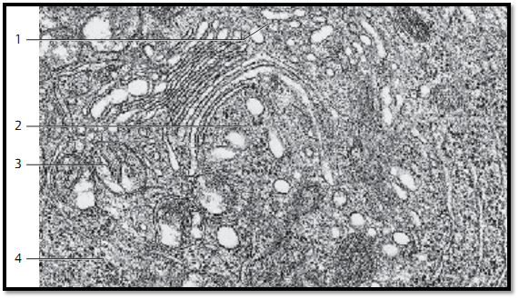
Exocrine cell from rat pancreas. Note the numerous vesicles of various sizes in the vicinity of the Golgi cisternae, and notice as well that the Golgi apparatus and the rough endoplasmic reticulum (rER) membranes 4 areina close spatial arrangement.
1 cis-Face 3 Mitochondria
2 trans-Face 4 Granular ER (rER)
This figure and next Fig. show that dictyosomes can have quite different shapes. The Golgi cisternae here are very bloated 1 .At the trans- face 2 are numerous vesicles, some are covered by a protein coat 3 . However, this coat is different from the well-known coat of “coated vesicles ,” which is made of clathrin. Section of an olfactory g land cell ( glandulae olfactoriae, Bowman glands).
1 Golgi cisternae 4 Granular ER (rER) membranes
2 trans-Face 5 cis-Face
3 Golgi vesicles with coat (protein cover)
This picture displays a mightily developed Golgi apparatus in an exocrine gland cell. It consists of several distended membrane cisternae 1 and shows on its concave side (trans-face ) 2 numerous Golgi vesicles of various sizes 3 and large, sometimes confluent secretor y vacuoles 4 . Section from a cell from the tissue of the Harder gland from Passer domesticus (sparrow).
1 Golgi cisternae
2 trans-Face
3 Golgi vesicles
4 Secretory vacuoles
5 cis-Face
References
Kuehnel, W.(2003). Color Atlas of Cytology, Histology, and Microscopic Anatomy. 4th edition . Institute of Anatomy Universitätzu Luebeck Luebeck, Germany . Thieme Stuttgar t · New York .


|
|
|
|
إجراء أول اختبار لدواء "ثوري" يتصدى لعدة أنواع من السرطان
|
|
|
|
|
|
|
دراسة تكشف "سببا غريبا" يعيق نمو الطيور
|
|
|
|
|
|
جمعية العميد تعقد اجتماعها الدوريّ لمناقشة أنشطتها العلمية المقبلة
|
|
|
|
قسم الشؤون الفكرية يقيم ندوةً عن أهمّية التداول المعرفي لوفدٍ من محافظة بابل
|
|
|
|
السيد الصافي يؤكد أن العتبات المقدسة تمتلك رؤية في دعم الكفاءات العراقية بمختلف تخصصاتها
|
|
|
|
استعدادًا لدوراته القرآنية الصيفية.. المجمع العلمي يقيم دورة تطويرية لمنتسبيه
|