آخر المواضيع المضافة

 النبات
الحيوان
الأحياء المجهرية
علم الأمراض
التقانة الإحيائية
التقنية الحيوية المكروبية
التقنية الحياتية النانوية
علم الأجنة
الأحياء الجزيئي
علم وظائف الأعضاء
الغدد
المضادات الحيوية
النبات
الحيوان
الأحياء المجهرية
علم الأمراض
التقانة الإحيائية
التقنية الحيوية المكروبية
التقنية الحياتية النانوية
علم الأجنة
الأحياء الجزيئي
علم وظائف الأعضاء
الغدد
المضادات الحيوية| Cardiac Muscle-Myocardium-Left Ventricle |


|
|
Read More
Date: 14-8-2016
Date: 27-7-2016
Date: 26-7-2016
|
Cardiac Muscle-Myocardium-Left Ventricle
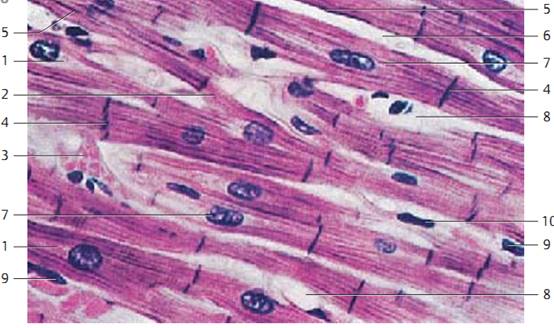
Like the skeletal musculature, the heart muscle tissue is striated. In contrast to skeletal muscles, cardiac muscle f ibers are branched and anastomose with each other, thus creating a network . The slit-shaped spaces in this web are filled with richly vascularize d connective tissue. Cardiac muscle f ibers are thinner than skeletal muscle fibers but thicker than smooth muscle cells. This figure clearly shows the capillaries 3 in the web spacing. The heavily stained nuclei are part of f ibrocytes 1 . Heart muscle cells ( cardiomyocytes ) have only one nucleus. Characteristic of cardiomyocytes are struts (disci in-tercalares ), which are dense, heavily stained bands across the muscle fibers. They are regularly spaced and cover the entire width of the fiber. Electron microscopic investigations have shown that struts are co-localize d with part of the cell borders. The faintly stained nuclei 2 occupy the center of the cardiac muscle cells. There are no fibrils in the perinuclear space. Instead, a rich layer of sarcoplasm surrounds the nuclei. In the sarcoplasmic cones are of ten fat droplets, glycogen and pigment granules (brown age pigment).
1 Fibrocyte
2 Nucleus of a hear t muscle cell
3 Capillary
Semi-thin section; stain: methylene blue-azure II; magnification: × 200
Cardiac Muscle—Myocardium—Left Ventricle
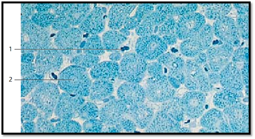
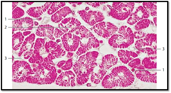
Cross-sections through cardiac muscle cells show the faintly stained central nuclei 2 and the Cohnheim fields or groups. The latter are base d on the formation of fibril bundles from myofibrils ( myofibril fields). The spaces between them contain sarcoplasm. Numerous capillaries 1 , which often still contain erythrocytes, are found in the loose connective tissue between muscle cells.
1 Capillaries
2 Nucleus of a cardiac muscle cell
Semi-thin section; stain: methylene blue-azure II; magnification: × 400
Cardiac Muscle—Myocardium—Left Ventricle
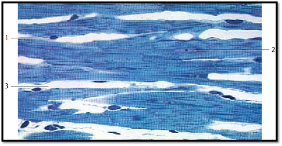
Longitudinal section through the myocardium of the lef t ventricle. Note the branching of the cardiomyocytes . The in part terrace-like arrange d struts 4 ( disci -intercalares ) are very noticeable because they stain heavily. Fibril-free sarcoplasm surrounds the nuclei like a cap 1 .
1 Perinuclear space
2 Branching of the cardiomyocytes
3 Vein with erythrocytes
4 Intercalated discs (disci -intercalares)
5 Network lamella of the cardiac muscle cell
6 Inter stitial connective tissue
7 Cardiac muscle cell with nucleus
8 Capillary
9 Endothelial nucleus
10 Nucleus of a fibrocyte
Stain: brilliant black-toluidine blue-safranin; magnification: × 200
Cardiac Muscle—Myocardium—Left Ventricle
This is another cross-section through the cardiac muscle tissue, which gives a clear picture of the Cohnheim grouping . Here, myofibrils have aggregate d to lamella in a radial formation. The sarcoplasm-rich spaces are free of fibrils and appear light. The size differences among nuclei should be noted 2 . In the sparse connective tissue between heart muscle cells are f ibrocytes and capillaries. It should also be noted that the apparent variations in the diameters of the cardiac muscle f ibers (cardiomyocytes ) depend on the presence or absence of their cell nuclei in the section.
1 Nucleus of a connective tissue cell
2 Nucleus of a cardiac muscle cell
3 Capillary
Stain: alum hematoxylin; magnification: × 600
Cardiac Muscle—Myocardium—Left Ventricle
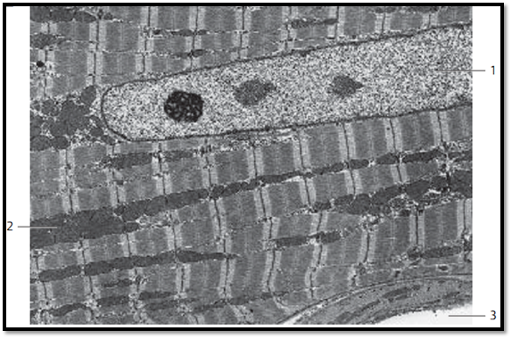
Section from a longitudinally cut cardiac muscle cell with central nucleus 1. The sarcomeres of the “cardiac muscle f ibers” have in principle the same striation pattern as the striate d skeletal muscle f ibers. However, in cardiac muscle sarcomeres are not arranged strictly parallel in separate columns, but form a branched three-dimensional web (cf. Figs. 238, 240). Note the mitochondria 2 , which are arrange d in columns between the microfilament bundles. In the nuclear cones are the sarcoplasm, mitochondria and, in numbers that increase with age, granules with the wear-and-tear pigment lipofuscin. In the lower right is a capillary 3 .
1 Nucleus of cardiomyocyte
2 Mitochondria
3 Capillary
Electron microscopy; magnification: × 2500
Cardiac Muscle—Myocardium—Left Ventricle
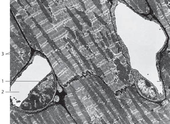
Section from several longitudinally cut cardiac muscle cells (cardiomyocytes ) after intravital staining by injection of ruthenium red. This dye clearly stains the intercellular space, the intercalate d discs 1 , and the pinocytotic vesicles of the endothelium. The struts follow an irregular, of ten terrace-like arrangement, which leads to a tight interlocking of the cells. Located at the inner surface of the cell membrane are electron-dense cadherins that allow actin filaments to attach and anchor themselves to this membrane. Therefore, the filaments do not bridge the intercellular space in the interlocking areas. On the contrary, there is a formation of desmosomes (maculae adherentes ) as well as focal adhesion centers, which are similar to the zonula adherentes and are name d fasciae adherentes. The mechanical transfer of the contraction forces between heart muscle cells takes place in these lo-cations.
1 Intercalated discs
2 Capillaris
3 Crist a-type mitochondria
Electron microscopy; magnification: × 2800
Cardiac Muscle—Myocardium—Left Ventricle
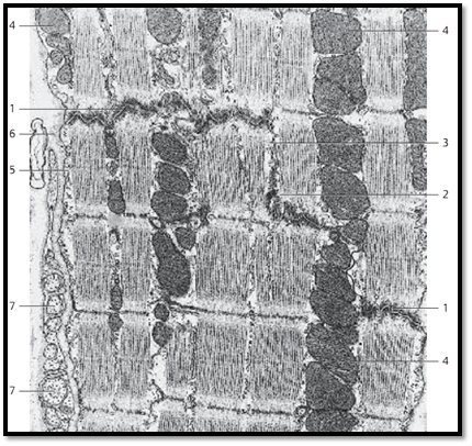
Data about the ultrastructure of heart muscle cells (cardiomyocytes ) indicate that the struts are arrange d in a terrace-like manner. This makes it possible to distinguish between transverse and longitudinal sectors. Inside the struts occur three different types of cell contacts: fascia adherentes 1 , maculae adherentes (desmosomes) 2 and nexus 3 . The fascia adherens exists as contact plate in the transverse sectors. There can b e single local desmosomes (maculae adherentes) as part of the fascia adherens or, as in this figure, in the longitudinal sectors of the struts. Gap junctions (nexus ) occur usually in the longitudinal sectors. They serve for electrical coupling between cardiac muscle cells and consist of the heart-specific nexus protein connexin 43 . Note the row of mitochondria with a large number of cristae 4.
Note: in contrast with skeletal musculature, the myocardium consists of separate single cells. The heart muscle tissue does not regenerate.
1 Fasciae adherentes
2 Macula adherens
3 Gap junction
4 Mitochondria
5 Plasmalemma with pinocytotic vesicles
6 Basal lamina
7 Axons without myelin sheath
Electron microscopy; magnification: × 30 000
References
Kuehnel, W.(2003). Color Atlas of Cytology, Histology, and Microscopic Anatomy. 4th edition . Institute of Anatomy Universitätzu Luebeck Luebeck, Germany . Thieme Stuttgar t · New York .


|
|
|
|
اكتشاف تأثير صحي مزدوج لتلوث الهواء على البالغين في منتصف العمر
|
|
|
|
|
|
|
زهور برية شائعة لتر ميم الأعصاب التالفة
|
|
|
|
|
|
المجمع العلمي يكرم القرّاء المشاركين في الختمات الرمضانية بقضائي المدائن والنهروان
|
|
|
|
العتبة العباسية تقيم ورشة علميّة عن عناصر الهُويّة الثقافيّة لوفدٍ من جامعة ذي قار
|
|
|
|
قسم الشؤون الفكرية يقيم ورشة تعريفية حول مشاريعه الرقمية في جامعة المثنى
|
|
|
|
العتبة العباسية تدعو للمشاركة في مسابقة القصّة القصيرة الثانية ضمن أسبوع الإمامة الدوليّ الثاني
|