آخر المواضيع المضافة

 النبات
الحيوان
الأحياء المجهرية
علم الأمراض
التقانة الإحيائية
التقنية الحيوية المكروبية
التقنية الحياتية النانوية
علم الأجنة
الأحياء الجزيئي
علم وظائف الأعضاء
الغدد
المضادات الحيوية
النبات
الحيوان
الأحياء المجهرية
علم الأمراض
التقانة الإحيائية
التقنية الحيوية المكروبية
التقنية الحياتية النانوية
علم الأجنة
الأحياء الجزيئي
علم وظائف الأعضاء
الغدد
المضادات الحيوية| Salmonella |


|
|
Read More
Date: 6-3-2016
Date: 3-3-2016
Date: 13-3-2016
|
Salmonella
Introduction
Salmonella is the main agent of foodborne diseases in several parts of the world (WHO, 2005). In the United States it is estimated that from two to four million cases of salmonellosis occur each year (FDA/CFSAN, 2009).
1.1 Taxonomic classification of Salmonella
Salmonella is a genus belonging to the family Enterobacteriaceae, defined by Brenner and Farmer III (2005) as rod-shaped, Gram-negative, non-spore-forming, facultative anaerobic and oxidase-negative bacteria. Both taxonomic classification and nomenclature are controversial, as summarized by Euzéby (2012a,b,c) and Grimont et al. (2000).
a) According to Grimont et al. (2000) the analysis of O and H antigens resulted in the description of a great number of Salmonella serovars over the years. Each serovar was considered as a species and names were given to more than 2000 serovars (e.g. “Salmonella london”). On the basis of biochemical reactions the genus Salmonella was divided by Kauff-mann into four subgenera designated by Roman numerals (I–IV), without formal nomenclature. Le Minor et al. (1970) considered Kauffmann’s sub-genera to represent species named ‘S. kauffmannii’ (subgenus I), ‘S. salamae’ (subgenus II), S. arizonae (subgenus III) and ‘S. houtenae’ (subgenus IV). Later, an additional subgroup (subgroup VI) was identified (Bongor group).
b) In 1980 the Approved Lists of Bacterial Names (Skerman et al., 1980) were published as a new starting point in bacterial nomenclature, including all the bacterial names having standing in nomenclature from 1 January 1980 (the names which did not appear in the lists lost standing in nomenclature and when cited should be printed with quotation marks). The genus Salmonella was included with five species: Salmonella choleraesuis, Salmonella enteritidis, Salmonella typhi, Salmonella typhimurium and Salmonella arizonae (Euzéby, 2012b).
c) Based on DNA relatedness studies Le Minor et al. (1982, 1986) considered all the Salmonella strains to constitute single species with seven subspecies: Salmonella choleraesuis subsp. arizonae, Salmonella choleraesuis subsp. bongori, Salmonella choleraesuis subsp. choleraesuis, Salmonella choleraesuis subsp. diarizonae, Salmonella choleraesuis subsp. houtenae, Salmonella choleraesuis subsp. indica and Salmonella choleraesuis subsp. salamae. Reeves et al. (1989) elevated the subspecies bongori in rank to the species Salmonella bongori. The name Salmonella bongori and the other six Salmonella choleraesuis subspecies names proposed by Le Minor have standing in nomenclature (Euzéby, 2012a,b).
d) Le Minor and Popoff (1987) submitted a request to the Judicial Commission of the International Committee on Systematics of Prokaryotes proposing to change the epithet choleraesusis to enterica in the name of the seven Salmonella subspecies, because cholerauis is also the name of a serovar. They also requested to consider the names Salmonella choleraesuis, Salmonella enteritidis, Salmonella typhi and Salmonella typhimurium as synonyms of Salmonella enterica subsp. enterica. The Judicial Commission decided negatively upon the request, prob-ably because it was not limited to nomenclature (the only scope of the Judicial Commission) and included the recognition of a single species in the genus Salmonella (a taxonomic proposal) (Grimont et al., 2000). However, the use of the non-validly published name Salmonella enterica spread among bacteriologists throughout the world (Euzéby, 2012b).
e) In 2005 the Judicial Commission of the International Committee on Systematics of Prokaryotes (2005) issued an opinion (Judicial Opinion 80) on some new requests that have been presentedas a result of the request of Le Minor and Popoff.
The Judicial Commission decided that the epithet enterica in Salmonella enterica should be conserved over all earlier epithets and that the subspecies and new combinations proposed by Le Minor and Popoff (1987) should be considered to be validly published. However, the Judicial Com-mission did not reject the epithet choleraesuis in Salmonella choleraesuis and the major problem following this decision is that now there are two systems of validly published names (Table.1): the “old” system (i.e. names validly published before publication of the Judicial Opinion 80, and the “new” system (i.e. names validly published as a consequence of the Judicial Opinion 80). The two systems can be used but the old is being used by an decreasing minority and new by an increasing majority (Euzéby, 2012b), including the World Health Organization Collaborating Center for Reference and Research on Salmonella, the Centers for Disease Control and Prevention (CDC) and the American Society for Microbiology (ASM) (Ellermeier and Slauch, 2005). Serovar names are no longer considered as species names and there-fore should not be printed in italics. Only serovars of S. enterica subsp. enterica are given names (usually geographical names). Serovars of other subspecies are designated by their O:H formula (White-Kauffmann-LeMinor antigenic formula) (Grimont et. al. (2000).
f ) Shelobolina et al. (2004) discovered a new species and proposed the name Salmonella subterranea, which was validly published in 2005. However, Euzèby (2012a) reported that, according to a personal communication from Shelobolina (June 05, 2010), this species is closely related to Escherichia hermannii and does not belong to the genus Salmonella.
Table.1 The two systems of validly published names of Salmonella (Euzéby, 2012a,b).
The strains most frequently involved in human diseases are S. enterica subsp. enterica, the habitat of which is the intestinal tract of hot-blooded animals and account for 99% of cases of salmonellosis in humans (Brenner et al., 2000). S. enterica subsp. salamae, subsp. arizonae and subsp. diarizonae are often isolated from the gut content of cold-blooded and rarely from humans or hot-blooded animals (Popoff and Le Minor, 2005). S. enterica subsp. houtenae and S. bongori are predominantly isolated from the environment and are rarely pathogenic to humans (Popoff and Le Minor, 2005, ICMSF, 1996).
1.2 Serological classification of Salmonella
The classification of Salmonella into species is little used in epidemiological studies, with the nomenclature linked to serotyping being much better known and more widely used. The extensively serotyping scheme used to determine a particular serovar is the White-Kauffmann-Le Minor identification system, based on the differences found in certain surface structures of the cells, which are antigenic. These structures are the cellular envelope or capsule (“Vi” capsular antigens), the cell wall (“O” somatic antigens) and the flagella (“H” flagellar antigens) (Ellermeier and Slauch, 2005, Popoff and Le Minor, 2005).
wall is composed of an inner layer of peptidoglycan, followed by an outer lipid-bilayer membrane (outer membrane) composed of lipoproteins, phospholipids and lipopolysaccharides (LPS). The LPS layer is divided into three portions, the internal lipid A portion, the core oligosaccharide region and the external repeating oligosaccharide chain, which forms the antigenic “O” region (O side-chain). The lipid A portion is the predominant responsible for the endotoxin effects of the LPS and the immune system of animals is exceedingly sensitive to it as a marker of infection. The “O” side chain (the O antigenic factor) is the serologically dominant part of the molecule. It is a repeated tetra or pentasaccharide, hydrophilic in nature, heat stable (100 or 120°C/2 h) and reaches out to the microenvironment of the bacterial cell (Rycroft, 2000). It may differ from one serovar to another in terms of the monosaccharides it contains, in the types of chemical bonds between these monosaccharides and in minor modifications, such as acetylation of the monosaccharides, for example (Ellermeier and Slauch, 2005).
Due to their importance to correct diagnosis, the factors are either classified as major or principal antigens or minor or secondary antigens. The major antigens serve as a basis to separate the Salmonella strains into somatic serogroups. For example, the somatic serogroup “A” includes all the strains containing the O:2 antigenic factor, which is not found in any other serogroup. The minor antigens are those that have a smaller discriminatory value and can be found in strains of more than one serogroup. For example, all the strains of the “A”, “B” and “D” serogroups have the O:12 antigenic factor, in addition to the factor that characterizes the group. The O groups were first designated by letters, but, when all the letters of the alphabet had been used, it was necessary to continue with numbers 51 to 67. The correspondence between the old designation (using letters) and the new designation (using numbers) is presented by Grimont et. al. (2007) and showed below:
Capsular (surface or envelope) antigens are very common in other Enterobacteriaceae genera (Escherichia coli and Klebsiella, for example), but are found in only few serovars of Salmonella. One specific capsular anti-gen is well-know, the Vi antigen, which occurs in three serovars of Salmonella: Typhi, Paratyphi C and Dublin. The strains of these serovars may or may not contain the Vi antigen, which, if present, masks the somatic (“O”) antigens and prevents agglutination with somatic antiserum. The inactivation of Vi antigem by heat (100ºC) allows the agglutination with the appropriate somatic antiserum (Popoff and Le Minor, 2005). The Vi antigen is the only true capsular polysaccharide produced by Salmonella spp. and was termed Vi because of its association with virulence (Rycroft, 2000).
Flagellar (“H”) antigens are derived from the flagella of motile strains. They are heat-sensitive and arise from variations in the amino acid sequence of the flagellar proteins. Most salmonellas have diphasic anti-genic flagellar expression, that is, a same strain has two genetic systems (genes distantly located on the chromosome) expressing different flagellins. Randomly and after 1000–10000 generations, the previously silent gene begins to be expressed and the other is silenced (phase variation). Some few serovars are monophasic, producing only one single type of flagellum (serovars Typhi and Enteritidis, for example, although atypical diphasic Salmonella Typhi have been isolated from Indonesia), while others are nonmotile, not producing any type of flagellum (serovars Pullorum and Gallinarum, for example) (Grimont et al., 2000, Grimont and Weill, 2007). The antigenic factors of phase one were originally designated by lowercase letters and, when all the letters of the alphabet had been used, the letter z, followed by a subscript number began to be used to identify the new factors. The antigens of phase two are designated by numbers, although a few strains produce antigens of phase one in phase two, which, in this case, are also identified by letters (Ellermeier and Slauch, 2005).
The White-Kauffmann-Le Minor System identifies and individualizes the Salmonella serovars by means of a formula composed of numbers and letters, which define the antigen(s) “O”, “Vi” and “H” present. The sequence is: 1st) the somatic antigens, 2nd) the Vi anti-gen, if present, and, between square brackets if their presence is not a constant in that particular serovar, 3rd) the flagellar antigens of phase one, if present, and 4th) the flagellar antigens of phase two, if present (Grimont and Weill, 2007). Examples: Serovar 9,12,[Vi]:d:- stands for major somatic factor O:9, minor somatic factor O:12, Vi may or may not be present (between parentheses), the phase-one flagellar antigen is d, and does have no phase-two flagellar anti-gen (monophasic). Serovar 1,4,[5],12:b:1,2 means: somatic factor O:1 underlined (indicating that it was originated by phage conversion), major somatic factor O:4, minor somatic factors O:5 (between square brackets indicating it may or may not be present) and O:12, the phase-one flagellar antigen is b and the phase-two flagellar antigens are 1 and 2.
More than 2,500 different Salmonella serovars had already been identified. The antigenic formula of each of these serovars is defined and maintained by the World Health Organization Collaborating Centre for Reference and Research on Salmonella at the Pasteur Institute, Paris, France (Grimont and Weill, 2007). Salmonella serovar nomenclature. The serovars of S. enterica subsp. enterica (S. choleraesuis subsp. choleraesuis) are also identified by names This is another source of confusion, since only two species are recognized in the genus Salmonella. According to Euzéby (2012b), the designation of the serovars is not regulated but it is advised to write them with capital letters and not in italic type: One of the following forms may be used: Salmonella London (shortened serovar nomenclature); Salmonella ser. London; Salmonella serovar London; Salmonella enterica subsp. enterica serovar London (complete name). The “World Health Organization’s International Center for Salmonella” and the “Centers for Disease Control and Prevention” use the shortened serovar nomenclature.
However, five serovars (Salmonella choleraesuis, Salmonella enteritidis, Salmonella paratyphi, Salmonella typhi and Salmonella typhimurium) have their names still validly published according to the old system of names before publication of the Judicial Opinion 80. Serovars most commonly found. In the 9th edition of the Antigenic Formulae of the Salmonella Serovars (Grimont and Weill, 2007) there are 2579 described serovars of Salmonella and more than 50% (1531) belong to S. enterica subsp. enterica, 505 belong to S. enterica subsp. salamae, 336 to S. enterica subsp. diarizonae, 99 to S. enterica subsp. arizonae, 73 to S. enterica subsp. houtenae, 13 to S. enterica subsp. indica and 22 to S. bongori. The most common somatic groups are A, B, C1, C2, D, E1 and E4 (Brenner et al., 2000, Ellermeier and Slauch, 2005). These serogroups account for about 99% of the Salmonella infections in humans and hot-blooded animals (Brenner et al, 2000), including serovars that are widely known, such as Paratyphi A (Group A), Paratyphi B and Typhimurium (Group B), Paratyphi C and Choleraesuis (Group C), Typhi, Enteritidis and Gallinarum (Group D). The antigenic factors found in these somatic serogroups are described by Grimont and Weill, 2007):
Group O:2 (A): Other O antigenic factors which can be found in strains of this group: O:1, O:12. Includes serovar Paratyphi A.
Group O:4 (B): Other O antigenic factors which can be found in strains of this group: O:1, O:5, O:12, O:27. Includes serovars Paratyphi B and Typhimurium.
Group O:7 (C1): Other O antigenic factors which can be found in strains of this group: O:6, O:14, Vi. Includes serovars Paratyphi C and Choleraesuis. The serovar containing the Vi antigen is Paratyphi C.
Group O:8 (C2-C3): Other O antigenic factors which can be found in strains of this group: O:6, O:20.
Group O:9 (D1): Other O antigenic factors which can be found in strains of this group: O:1, O:12, Vi. Includes serovars Typhi, Enteritidis and Gallinarum. The serovars containing the Vi antigen are Typhi and Dublin.
Group O:9,46 (D2): Other O antigenic factors which can be found in strains of this group: O:1.
Group O:9,46,27 (D3): Other O antigenic factors which can be found in strains of this group: O:1, O:12.
Group O:3,10 (E1): Other O antigenic factors which can be found in strains of this group: O:15, O:34.
Group O:1,3,19 (E4): Other O antigenic factors which can be found in strains of this group: O:10, O:15.
1.3 Biochemical characteristics of Salmonella
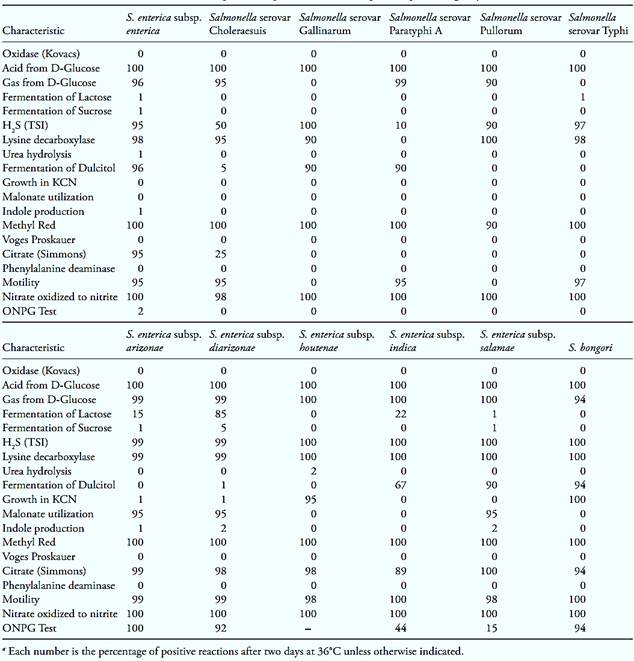
According to Popoff and Le Minor (2005) the main biochemical characteristics of the species and subspecies of Salmonella and of some important serovars are summarized in Table 2. The salmonellas, like all Enterobacteriaceae, are oxidase-negative. They do also no produce butylene glycol (Voges-Proskauer-negative), nor phenylalanine deaminase. They are normally motile, although strains of the Gallinarum and Pullorum serovars are usually nonmotile. They reduce nitrate to nitrite (except for 2% of the strains of the Choleraesuis serovar) and are MR (Methyl Red) positive (except for 9% of the strains of the Pullorum serovar). They normally produce gas from glucose, but the strains of the Typhi and Gallinarum serovars test negative for this fermentation reaction.
According to ICMSF (1996) the temperature growth range varies from 5–7°C to 46°C, with the optimal temperature for growth ranging between 35 and 43°C. The growth pH varies from 3.8 to 9.5, with an optimum in the 7.0 to 7.5 range. The minimum water activity for growth is 0.94.
The strains of Salmonella enterica subsp. enterica are the main target in the analysis of foods, with the bio-chemical profile described below (data from Popoff and Le Minor, 2005) being commonly considered as typical in detection tests.
a) Ferment glucose with the production of acid (100% of the strains) and gas (96% of the strains).
b) Do not ferment lactose and sucrose (99% of the strains).
c) Decarboxylate lysine (98% of the strains) with the exception of serovar Paratyphi A (100% of the strains are negative for this characteristic).
d) Produce H2S (95% of the strains), with the exception of serovars Paratyphi A (90% of the strains negative) and Choleraesuis (50% of the strains negative).
e) Do not produce urease (99% of the strains).
f ) Ferment dulcitol (96% of the strains), with the exception of serovars Typhi (100% of the strains negative), Pullorum (100% of the strains negative) and Choleraesuis (95% of the strains negative).
g) Do not grow in the presence of KCN (100% of the strains).
h) Do not use malonate (100% of the strains).
i) Use citrate (95% of the strains), with the exception of the serovars Typhi, Paratyphi A, Pullorum Gallinarum (100% of the strains negative) and Choleraesuis (75% of the strains negative).
j) Do not produce indole (99% of the strains).
k) Do not produce the β-galactosidase enzyme, negative for the ONPG test (ortho-Nitrophenyl-β-D- galactopyranoside (98% of the strains).
Table 2 Biochemical reactions of Salmonella species, subspecies and serovars important epidemiologically (Brenner and Farmer III, 2005)a.
1.4 Epidemiology
The principal habitat of the salmonellas is the intestinal tract of humans and animals. Some few serovars are can be found predominantly in one particular host, such as Salmonella Typhi, Salmonella Paratyphi A and C and Salmonella Sendai that are strictly human, Salmonella Abortusovis in ovines, Salmonella Abortusequi in equines, Salmonella Gallinarum in poultry, Salmonella Typhisuis in swine, Salmonella Choleraesuis in swine (more rarely, in humans) and Salmonella Dublin in bovine (more rarely in humans and ovines) (Ellermeier and Slauch, 2005). When these serovars cause diseases in humans, the process is generally invasive and is life threatening (WHO, 2005).
Most serovars, however, have a wide spectrum of hosts and typically cause gastroenteritis without complications which do not require special treatment. In children, the elderly, and debilitated or immunologically compromised individuals, on the other hand, these infections can be severe. The most important serovars involved in transmitting these salmonelloses from animals to humans are Salmonella Enteriditis and Salmonella Typhimurium (WHO, 2005).
According to FDA (2012) Salmonella Typhi and Paratyphi cause typhoid fever in humans, which occurs between one to three weeks, but may be as long as 2 months after exposure. The infective dose is fewer than 1,000 cells and the symptoms include high fever, lethargy, abdominal pains, diarrhea or constipation, headache; achiness, loss of appetite and, sometimes, a rash of rose-colored spots. Duration is generally 2 to 4 weeks. Complications may occur, including septicemia and septic arthritis. Chronic infection of the gallbladder may occur, causing the infected person to become a carrier. The mortality rate of typhoid fever (untreated) is 10%.
The gastroenteritis caused by other salmonellas occur between six and 72 hours, including fever, headache, abdominal colic, diarrhea, nausea and vomiting. Dura-tion varies from four to seven days (one to two days for the acute symptoms) but may be prolonged depending on the host, the dose ingested and the Salmonella strain involved. The infective dose may be very low (one cell) depending on the host and strain involved. The illness is generally self-limiting among healthy people with intact immune systems and the mortality is generally less than 1%. The very young, the elderly, the AIDS or chronic illnesses patients and the people using medications for cancer (chemotherapy) or immunosuppressive drugs are more vulnerable. AIDS patients are affected by salmonellosis with a frequency that is twenty-fold greater than that in the general population, suffering from recurrent episodes (FDA/CFSAN, 2012).
The usual sources of Salmonella Typhi and Salmonella Paratyphi in the environment are drinking and/or irrigation water contaminated by untreated sewage. Other salmonellas are widespread among animals and are spread through the fecal-oral route, reaching the natural environment (water, soil, insects) and contaminating meat, produce in the field, factory equipment, hands, and kitchen surfaces and utensils. The disease is generally contracted through the consumption of contaminated foods of animal origin (meats, poultry, eggs, milk and dairy products, fish, shrimp) but fresh pro-duce (fruit and vegetables such tomatoes, peppers, and cantaloupes) and low-moisture foods (such as spices) also have been implicated in transmission. The bacteria reaches the entire food production chain, from primary products onwards, and other foods that already have been implicated in the transmission of salmonelloses include yeast, coconut, sauces, salad dressings, cake mixes, cream-filled desserts and toppings, dried gelatin, peanut butter, cocoa, and chocolate. Cross contamination is another common form of food contamination with Salmonella (FDA/CFSAN, 2012).
1.5 Traditional methods used for the examination of Salmonella
The traditional detection technique used to detect Salmonella in foods is a classical culture presence/absence method, specifically developed to ensure the detection even in the most extremely unfavorable situations. This is the case of foods containing a competing microbiota that is much larger than the Salmonella population and/or foods in which the Salmonella cells are present in only very low numbers and/or foods that contain cells injured by preservation processes (heat treatments, freezing, drying). The procedures recommended by the different regulatory authorities, though they show some variations in the selection of the culture media and the way in which the samples are to be prepared, all basically follow four steps that can be applied to any type of food. These steps and the media used by the different international regulatory organizations are summarized in Table 3.
Pre-enrichment in non-selective broth: The objective of this step is to recover injured cells, which can be obtained by incubating the sample in non-selective conditions, for at least 18 hours. The most commonly used media are Buffered Peptone Water (BPW) and Lactose Broth (LB), with some exceptions presented in the description of the procedures.
Enrichment in selective broth: The objective of this step is to inhibit the multiplication of the accompanying microbiota and preferentially promote the increase of the number of Salmonella cells, by incubating the pre-enriched sample in selective broth, for 18 to 24 hours. In this step, it is recommended that two different enrichment media be used, since the resistance of Salmonella to selective agents varies from strain to strain. The most recommended media for this purpose are Modified Rappaport-Vassiliadis Broth (RV) or Rappaport-Vassiliadis Soy Broth (RVS) and diverse formulations of Tetrathionate Broth. The selectivity of RV and
Table 3 Media and incubation conditions recommended by ISO, FDA and USDA methods for Salmonella in foods.
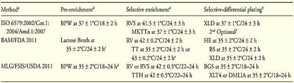
a ISO 6579:2002/Cor.1:2004/Amd.1:2007 = Method of International Organization for Standardization, BAM/FDA 2011 = Method of Bacteriological Analytical Manual Online, Food and Drug Administration (Andrews and Hammack, 2011), MLG/FSIS/USDA 2011 = Method of Microbiological Laboratory Guidebook Online, Food Safety and Inspection Service, United States Department of Agriculture (MLG/FSIS/USDA, 2011).
b BPW = Buffered Peptone Water, BGS = Brilliant Green Sulfa Agar, BS = Bismute Sulfite Agar, DMLIA = Double Modified Lysine Iron Agar, HE = Hektoen Enteric Agar, MKTTn = Muller Kauffmann Tetrathionate Novobiocin Broth, RV = Modified Rappaport Vassiliadis Broth, RVS = Rappaport-Vassiliadis Soya Broth, TT = Tetrathionate Broth, TTH = Tetrathionate Broth Hajna, XLD = Xylose Lysine Desoxycholate Agar, XLT4 = Xylose Lysine Tergitol 4 Agar.
c There are variations presented in the descriptions of the procedure and in the Table 19.6.
d There are variations in the incubation time, presented in the descriptions of the procedure.
e 35°C for foods with a low microbial load and 43°C for foods with a high microbial load.
f ISO recommends a medium complementary to XLD for lactose positive Salmonella, Salmonella Typhi and Salmonella Paratyphi strains. Bismuth Sulfite Agar (BS) and Brilliant Green Agar (BG) can be used.
RVS is based on the presence of malachite green oxalate, on high osmotic pressure (presence of a high concentration of magnesium chloride) and on the relatively acidic pH (5.1). There are several commercial formulations of Tetrathionate Broth available, which are used by different regulatory organizations. These formulations are well defined in the description of the procedures. The basis of selectivity of the medium is the presence of brilliant green, bile (or sodium deoxycholate), iodine and sodium thiosulfate. Iodine is added at the time of use, and reacts with the thiosulfate forming tetrathionate. The growth of enterobacteria that reduce tetrathionate, such as Salmonella, is relatively normal, while the coliforms and other non-reducing Enterobacteriaceae are suppressed. The reduction of tetrathionate produces acid, neutralized by calcium carbonate contained in the formulation. Proteus is also reducers of tetrathionate and may grow in this medium, an interference that can be reduced with the addition of novobiocin.
Differential selective plating: The objective of this step is to preferentially promote the development of Salmonella colonies exhibiting typical characteristics that distinguish them from competitors, for subsequent serological and biochemical confirmation. Just like as for the selective enrichment step, it is recommended that differential plating be done on more than one type of culture medium, and there are several media available for use in this step. The most commonly used are media that differentiate Salmonella through its incapacity to ferment lactose and the concomitant ability to produce H2S, such as Hektoen Enteric Agar (HE), Xylose Lysine Desoxicolate Agar (XLD) and Xylose Lysine Tergitol 4 Agar (XLT4). Since there are Salmonella strains that ferment lactose or do not produce H2S, it is important that the second or third plating medium be not based on any of these two characteristics. One of these options is Brilliant Green Agar (BG), which is based on the fermentation of lactose but not on the production of H2S, and Bismuth Sulfite Agar (BS), which is based on the pro-duction of H2S, but not on the fermentation of lactose.
Confirmation: The objective of this step is to verify whether the colonies obtained on the plates are actually Salmonella colonies, by means of biochemical and serological assays. Biochemical confirmation aims at verifying the characteristic biochemical profile of strains of Salmonella enterica subsp. enterica presented in Table 2. In general, the different regulatory organizations or authorities also recommend the use of commercial miniaturized kits, which allow performing a greater number of biochemical tests. Serological confirmation verifies the presence of the “O”, “Vi” and “H” antigens, using agglutination tests with polyvalent antisera. These antisera should contain antibodies for the factors most commonly encountered and which, in the case of the somatic serological test, belong to the serogroups “A” to “E”. Some commercial antisera contain also antibodies for the Vi antigen. Some brands include a complete pool, containing antibodies for the somatic A, B, C1, C2, D, E1 and E4 groups, the flagellar and the Vi antigens, all together. The methods of food analysis conclude confirmation at this point, since complete characterization of Salmonella strains is normally done by only a few reference laboratories in each country.
1.6 Alternative methods for the analysis of Salmonella
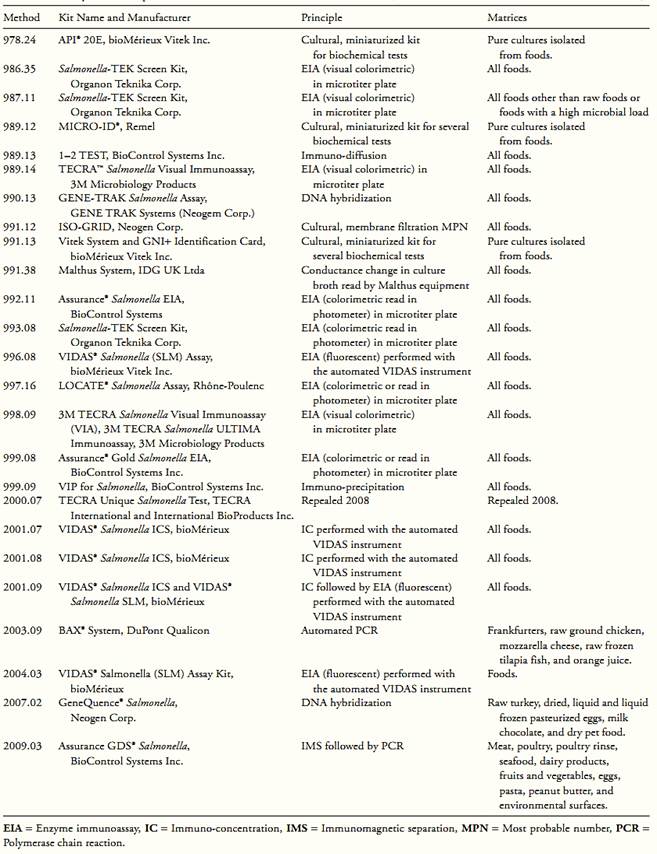
The traditional method is quite sensitive, with a detection limit of one colony forming unit/25 g of sample, but is slow and laborious. Because of this, there is great interest in rapid and simpler methods that might be used instead of the traditional method. Over the past ten years, great advances have been made in the development of new methods, particularly immunological methods and methods based on nucleic acids. These methods follow the current trend of developing registered trademarked analytical test kits, defined by the AOAC International as: “systems containing all key components required to conduct the examination of one or more microorganisms, in one or more foods, in accordance with a certain method” (Andrews, 1997).
The great advantage of these kits is that the all the materials necessary to perform the tests (partially or in their entirety) are marketed together, eliminating preparation in the laboratory. Alternative methods that already have been officially recognized by the AOAC International are the micro-biological test kits described in Table 4.
1.7 Composite samples for analysis
In the microbiological examination of several separate samples units of a product lot, whenever there is evidence that the composition will not affect the result for that type of food, the practice of pooling the sam-ples may be utilized. BAM/FDA (Andrews and Hammack, 2011) specifies that up to 15 sample units may be pooled into a composite sample, with the exceptions shown in Table 6. The methods MLG/FSIS/USDA 2011 and ISO 6579:2002/Cor.1:2004/Amd.1:2007 do not specify a limit relative to the maximum number of sample units that may be pooled to form a composite sample. There are two procedures that can be used for pooling samples:
Pooling before the pre-enrichment: Consists in collecting one analytical unit (generally 25 g) of each of the sample units and then pooling all analytical units collected together into one single composite sample, mixing well the entire content. To this composite sample, the pre-enrichment broth is added in an amount sufficient to obtain a 10−1 dilution. For example, to pool 10 × 25 g, 2,250 ml of pre-enrichment broth is used. When the volume of pre-enrichment broth is very large, pre-heat the broth to the incubation temperature prior the inoculation. This type of pooling is recommended for samples in which absence of Salmonella is expected or, if the microorganism be present, it is not important to determine exactly which sample unit is contaminated. This procedure is not recommended for foods that do not allow obtaining a homogeneous composite sample.
Pooling after the pre-enrichment: Consists in separately pre-enrich each analytical unit first and then pooling the pre-enrichment broth together into composite pre-enrichment broths. In this case, the volume of the selective enrichment broth must be large enough to maintain the proportion recommended for the pre-enrichment to be transferred. For example, 0.1 ml of pre-enrichment normally is transferred to 10 ml of Rappaport-Vassiliadis (RV or RVS) Broth. Thus, in order to pool together 10 samples (0.1 ml of each pre-enriched broth), 100 ml of RV or RVS is necessary. When the volume of selective enrichment broth is very large, pre-heat the broth to the incubation temperature prior the inoculation. This type of pooling samples is recommended for foods that do not allow obtaining a homogenous sample by pooling before the pre-enrichment, or also when it is important to determine exactly which sample unit is contaminated, if Salmonella is detected. In this situation, it is possible to analyze individually each pre-enriched unit, preserved under refrigeration.
Table.4 Analytical kits adopted as AOAC Official Methods for Salmonella in foods (Horwitz and Latimer, 2010, AOAC International, 2010).
References
Andrews, W.H. (1997) New trends in food microbiology: an AOAC International perspective. Journal of AOAC International, 80(4), 908–912.
Andrews, W.H. & Hammack, T.S. (2011) Salmonella. In: FDA (ed.) Bacteriological Analytical Manual, Chapter 5. [Online] Silver Spring, Food and Drug Administration. Available from: http:// www.fda.gov/Food/ScienceResearch/LaboratoryMethods/Bacte-riologicalAnalyticalManualBAM/ucm070149.htm [accessed in 10th February 2012].
AOAC International (2010). Rapid Methods Adopted as AOAC Official MethodsSM. [Online] Available from: http://www.aoac.org/vmeth/oma_testkits.pdf [Accessed 26th April 2011).
Brenner, D.J. & Farmer III, J.J. (2005) Family I. Enterobacteriaceae. In: Brenner, D.J., Krieg, N.R. & Staley, J.T. (eds). Bergey’s Manual of Systematic Bacteriology. Volume 2. 2nd edition. New York, Springer Science+Business Media Inc. pp. 587–607.
Brenner, F.W., Villar, R.G., Angulo, F.J., Tauxe, R. & Swaminathan, B. (2000) Salmonella nomenclature. Journal of Clinical Microbiology, 38(7), 2465–2467.
Ellermeier, C.D. & Slauch, J.M. (2005) The Genus Salmonella. In: Dworkin, M., Falkow, S., Rosenberg, E., Schleifer, K.H. & Stackebrandt, E. (eds). The Prokaryotes: An evolving electronic resource for the microbiological community. 3rd edition, Release 3.20, 12/31/2005 [Online]. New York, Springer-Verlag. Avail-able from: http://141.150.157.117:8080/prokPUB/index.htm [Accessed 1st March 2006].
Euzéby, J.P. (2012a) List of Prokaryotic names with Standing in Nomenclature – Genus Salmonella. [Online] Available from: http://www.bacterio.cict.fr/s/salmonella.html [Accessed 8th August 2012].
Euzéby, J.P. (2012b) List of Prokaryotic names with Standing in Nomenclature – Salmonella nomenclature. [Online] Availabe from: http://www.bacterio.cict.fr/salmonellanom.html [Accessed 8th August 2012].
Euzéby, J.P. (2012c) List of Prokaryotic names with Standing in Nomenclature – Approved Lists of Bacterial Names. [Online] Availabe from: http://www.bacterio.cict.fr/alintro.html [Accessed 8th August 2012].
FDA/CFSAN (ed.) (2012) Foodborne Pathogenic Microorganisms and Natural Toxins Handbook “Bad Bug Book”. 2nd edition. [Online] Silver Spring, Food and Drug Administration, Center for Food Safety & Applied Nutrition. Available from: http://www.fda.gov/food/foodsafety/foodborneillness/foodborneillnessfoodbornepa-thogensnaturaltoxins/badbugbook/default.htm [Accessed 10th July 2012].
Grimont, P.A.D., Griomont, F. & Bouvet, P. (2000) Taxonomy of the Genus Salmonella. In: Wray, C & Wray A. (eds) Salmonella in Domestic Animals. [Online] London, CAB International. Available from: http://cremm.es/ARTICULOS/Salmonella%20 in%20Domestic%20Animals.pdf [Accessed 10th July 2012]. pp. 1–17.
Grimont, P.A.D. & Weill, F-X. (2007) Antigenic Formulae of the Salmonella Serovars. 9th edition. [Online] Paris, France, World Health Organization Collaborating Centre for Reference and Research on Salmonella. Available from: http://nih.dmsc.moph.go.th/aboutus/media/antigenic%20formula%20of%20Salmonella.pdf [Accessed 14th August 2012].
FDA/CFSAN (ed.) (2009) Foodborne Pathogenic Microorganisms and Natural Toxins Handbook “Bad Bug Book”. [Online] College Park, Food and Drug Administration, Center for Food Safety & Applied Nutrition. Available from: http://www.fda.gov/Food/FoodSafety/FoodborneIllness/FoodborneIllnessFoodbornePa-thogensNaturalToxins/BadBugBook/ucm069966.htm [accessed 1st November 2011].
Horwitz, W. (ed.) (2000) Official Methods of Analysis of AOAC International. 17th edition. Gaithersburg, Maryland, AOAC International.
Horwitz, W. & Latimer, G.W. (eds) (2010) Official Methods of Anal-ysis of AOAC International. 18th edition., revision 3. Gaithersburg, Maryland, AOAC International.
ICMSF (International Commission on Microbiological Specifications for Foods) (1996) Microorganisms in Foods 5. Microbiological Specifications of Food pathogens. London, Blackie Academic & Professional.
International Organization for Standardization (2007) ISO 6579:2002/Cor 1:2004/Amd 1:2007. Microbiology of food and animal feeding stuffs – Horizontal method for the detection of Sal-monella spp. 4th edition: 2002, Corrigendum 1:2004, Amend-ment 1:2007. Geneva, ISO.
Judicial Commission of The International Committee on Systematics of Prokaryotes (2005) The type species of the genus Salmonella Lignieres 1900 is Salmonella enterica (ex Kauffmann and Edwards 1952) Le Minor and Popoff 1987, with the type strain LT2T, and conservation of the epithet enterica in Salmonella enterica over all earlier epithets that may be applied to this species. Opinion 80.
International Journal of Systematic and Evolutionary Microbiology, 55, 519–520.
Le Minor, L.E (1984) Genus III Salmonella Lingered. In: Krieg, N.R. & Holt, J.G. (eds). Bergey’s Manual of Systematic Bacteriology. Volume 1, 1st edition. Baltimore, Williams & Wilkins. pp. 427–458.
Le Minor, L. & Popoff, M.Y. (1987) Request for an Opinion. Designation of Salmonella enterica sp. nov., nom. rev., as the type and only species of the genus Salmonella. International Journal of Systematic and Evolutionary Microbiology, 37, 465–468.
Le Minor, L., Popoff, M.Y., Laurent, B. & Hermant, D. (1986) Individualisation d’une septième sousespèce de Salmonella: S. choleraesuis subsp. indica subsp. nov. Annales de l’Institut Pasteur/Microbiologie 137B, 211–217.
Le Minor, L., Rohde, R. & Taylor, J. (1970) Nomenclature des Sal-monella. Annales de l’Institut Pasteur 119, 206–210. Le Minor, L., Veron, M. & Popoff, M.Y. (1982) Taxonomie des Salmonella. Annales de Microbiologie 133B, 223–243.
MLG/FSIS/USDA (2008) Most probable number procedure and tables. In: Microbiology Laboratory Guidebook [Online] Washington, Food Safety and Inspection Service, United States Department of Agriculture. Available from: http://www.fsis.usda.gov/PDF/MLG_Appendix_2_03.pdf [Accessed 3rd November 2011].
MLG/FSIS/USDA (2011) Isolation and Identification of Salmonella from Meat, Poultry, Pasteurized Egg and Catfish Products. In: Microbiology Laboratory Guidebook [Online] Washington, Food Safety and Inspection Service, United States Department of Agriculture. Available from: http://www.fsis.usda.gov/PDF/MLG_4_05.pdf [Accessed 10th February 2012].
Popoff, M.Y. & Le Minor, L.E., 2005. Genus XXXIII Samonella. In: Brenner, D.J., Krieg, N.R. & Staley, J.T. (eds). Bergey’s Man-ual of Systematic Bacteriology. Volume 2. 2nd edition. New York, Springer Science+Business Media Inc. pp. 764–799.
Reeves, M.W., Evins, G.M., Heiba, A. A., Plikaytis, B.D. & Farmer III, J.J. (1989) Clonal nature of Salmonella typhi and its genetic relatedness to other salmonellae as shown by multilocus enzyme electrophoresis and proposal of Salmonella bongori comb. nov. Journal of Clinical Microbiology, 27, 313–320.
Rycroft, A.N. (2000) Structure, function and synthesis of surface polysaccharides in Salmonella. In: Wray, C & Wray A. (eds) Salmonella in Domestic Animals. [Online] London, CAB International. Available from: http://cremm.es/ARTICULOS/Sal-monella%20in%20Domestic%20Animals.pdf [Accessed 10th July 2012]. pp. 19–33.
Shelobolina, E.S., Sullivan, S.A., O’Neill, K.R., Nevin, K.P. & Lovley, D.R. (2004) Isolation, characterization, and U(VI)-reducing potential of a facultatively anaerobic, acid-resistant bacterium from low-pH, nitrate- and U(VI)-contaminated subsurface sediment and description of Salmonella subterranea sp. nov. Applied and Environmental Microbiology, 70, 2959–2965.
Skerman, V.B.D., McGowan, V. & Sneath, P.H.A. (1980) Approved Lists of Bacterial Names. International Journal of Systematic Bacteriology, 30, 225–420.
WHO (World Health Organization). (2005) Drug-resistant Salmonella. Fact Sheet N°139. [Online] Available from: http://www.who.int/mediacentre/factsheets/fs139/en/ [Accessed 1st November 2011].


|
|
|
|
"وجه أوزمبيك".. تحذير من عرض غير متوقع لدواء إنقاص الوزن
|
|
|
|
|
|
|
"واتساب" يتوقف عن العمل في 3 هواتف شهيرة.. هل تمتلك أحدها؟
|
|
|
|
|
|
|
مدينة الفردوس الترفيهية توفر أكثر من 60 جلسة عائلية عامة وخاصة
|
|
|