آخر المواضيع المضافة

 النبات
الحيوان
الأحياء المجهرية
علم الأمراض
التقانة الإحيائية
التقنية الحيوية المكروبية
التقنية الحياتية النانوية
علم الأجنة
الأحياء الجزيئي
علم وظائف الأعضاء
الغدد
المضادات الحيوية
النبات
الحيوان
الأحياء المجهرية
علم الأمراض
التقانة الإحيائية
التقنية الحيوية المكروبية
التقنية الحياتية النانوية
علم الأجنة
الأحياء الجزيئي
علم وظائف الأعضاء
الغدد
المضادات الحيوية| RNA Editing Can Be Directed by Guide RNAs |


|
|
Read More
Date: 17-11-2020
Date: 10-12-2015
Date: 2-5-2016
|
RNA Editing Can Be Directed by Guide RNAs
KEY CONCEPTS
- Extensive RNA editing in trypanosome mitochondria occurs by insertions or deletions of uridine.
- The substrate RNA base pairs with a guide RNA on both sides of the region to be edited.
- The guide RNA provides the template for addition (or less often, deletion) of uridines.
- Editing is catalyzed by the editosome, a complex of endonuclease, exonuclease, terminal uridyl transferase activity, and RNA ligase.
Another type of editing is revealed by dramatic changes in sequence in the products of several genes of trypanosome mitochondria. In the first case discovered, the sequence of the cytochrome oxidase subunit II protein has an internal frameshift that is not predicted based on the nucleotide sequence of the coxII gene. The sequences of the gene and protein given in FIGURE 1 are conserved in several trypanosome species, thus the method of RNA editing is not unique to a single organism.
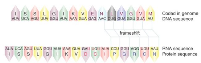
FIGURE 1. The mRNA for the trypanosome coxII gene has a frameshift relative to the DNA; the correct reading frame observed in the protein is created by the insertion of four uridines (shown inred).
The discrepancy between the sequence of the coxII gene and the protein product is due to an RNA-editing event. The coxII mRNA has an insert of an additional four nucleotides (all uridines) around the site of frameshift. The insertion establishes the proper readingfra me for the protein. No second coxII gene carrying the frameshift sequence has been discovered, so we are forced to conclude that the extra bases are inserted during or after transcription. A similar discrepancy between mRNA and genomic sequences is found in genes of the SV5 and measles paramyxoviruses, in these cases involving the addition of G residues in the mRNA.
Similar editing of RNA sequences occurs for other genes and includes deletions as well as additions of uridine. The extraordinary case of the cytochrome c oxidase III (coxIII) gene of Trypanosoma brucei is summarized in FIGURE 2. More than half of the residues in the mRNA consist of uridines that are not encoded by the gene. Comparison between the genomic DNA and the mRNA shows that no stretch longer than seven nucleotides is represented in the mRNA without alteration, and runs of uridine up to seven bases long are inserted. The information for the specific insertion of uridines is provided by a guide RNA.
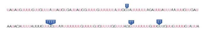
FIGURE 2. Part of the mRNA sequence of T. brucei coxIII shows many uridines that are not encoded in the DNA (shown in red) or that are removed from the RNA (shown as Ts in blue boxes).
Guide RNA contains a sequence that is complementary to the correctly edited mRNA. FIGURE 3 shows a model for its action in the cytochrome b gene of another trypanosome,
Leishmania. The sequence at the top of the figure shows the original transcript, or pre-edited RNA. Gaps show where bases will be inserted in the editing process. Eight uridines must be inserted into this region to result in the final mRNA sequence. The guide RNA is complementary to the mRNA for a significant length, including and surrounding the edited region. Typically the complementarity is more extensive on the 3′ side of the edited region and is rather short on the 5′ side. Pairing between the guide RNA and the pre-edited RNA leaves gaps where unpaired A residues in the guide RNA do not find complements in the preedited RNA. The guide RNA provides a template that allows the missing U residues to be inserted at these positions in a process described in the next paragraph. When the reaction is completed the guide RNA separates from the mRNA, which becomes available for translation.
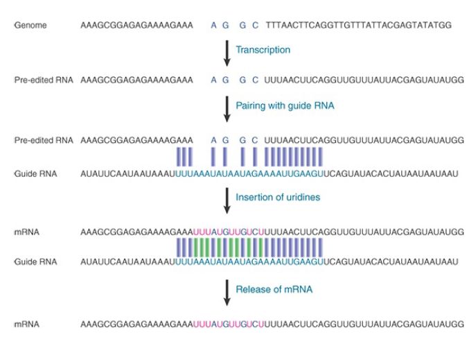
FIGURE 3. Pre-edited RNA base pairs with a guide RNA on both sides of the region to be edited. The guide RNA provides a template for the insertion of uridines. The mRNA produced by the insertions is complementary to the guide RNA.
Specification of the final edited sequence can be quite complex. In the example of Leishmania cytochrome b, a lengthy stretch of the transcript is edited by the insertion of a total of 39 U residues, which appears to require two guide RNAs acting at adjacent sites. The first guide RNA pairs at the 3′-most site, and the edited sequence then becomes a substrate for further editing by the next guide RNA. The guide RNAs are encoded as independent transcription units. FIGURE 4 shows a map of the relevant region of the Leishmania mitochondrial DNA. It includes the gene for cytochrome b, which encodes the pre-edited sequence and two regions that specify guide RNAs. Genes for the major coding regions and for their guide RNAs are interspersed.
In principle, a mutation in either the gene or one of its guide RNAs could change the primary sequence of the mRNA, and thus the primary sequence of the polypeptide. By genetic criteria, each of these units could be considered to comprise part of the gene. The units are independently expressed, and as a result they should complement in trans. If mutations were available, three complementation groups would be needed to encode the primary sequence of a single protein.
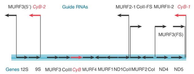
FIGURE 4. The Leishmania genome contains genes encoding pre-edited RNAs interspersed with units that encode the guide RNAs required to generate the correct mRNA sequences. Some genes have multiple guide RNAs. CyB is the gene for pre-edited cytochrome b, and CyB-1 and CyB-2 are genes for the guide RNAs involved in its editing.
The characterization of intermediates that are partially edited suggests that the reaction proceeds along the pre-edited RNA in the 3′–5′ direction. The guide RNA determines the specificity of uridine insertions by its pairing with the pre-edited RNA. Editing of uridines is catalyzed by a 20S enzyme complex called the editosome that is composed of about 20 proteins and contains an endonuclease, a terminal uridyl transferase (TUTase), a 3′–5′ Uspecific exonuclease (exoUase), and an RNA ligase. As illustrated in FIGURE 5, the editosome binds the guide RNA and uses it to pair with the pre-edited mRNA. The substrate RNA is cleaved at a site that is presumably identified by the absence of pairing with the guide RNA; a uridine is inserted or deleted to base pair with the guide RNA, and then the substrate RNA is ligated. Uridine triphosphate (UTP) provides the source for the uridyl residue. It is added by the TUTase activity. Deletion of U residues is mediated by an exoUase, which functions in concert with a 3′ phosphatase to allow the newly edited RNA construct to religate.
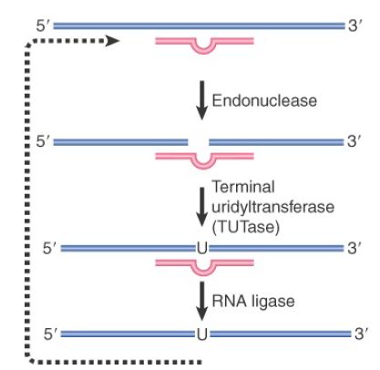
FIGURE 5. Addition or deletion of U residues occurs by cleavage of the RNA, removal or addition of the U, and ligation of the ends. The reactions are catalyzed by a complex of enzymes under the direction of guide RNA (red line).
The structures of partially edited molecules suggest that the U residues are added one at a time rather than in groups. It is possible that the reaction proceeds through successive cycles in which U residues are added, tested for complementarity with the guide RNA, retained if acceptable, and removed if not, so that the construction of the correct edited sequence occurs gradually. We do not know whether the same types of reaction are involved in editing reactions that add C residues.


|
|
|
|
إجراء أول اختبار لدواء "ثوري" يتصدى لعدة أنواع من السرطان
|
|
|
|
|
|
|
دراسة تكشف "سببا غريبا" يعيق نمو الطيور
|
|
|
|
|
|
لأعضاء مدوّنة الكفيل السيد الصافي يؤكّد على تفعيل القصة والرواية المجسّدة للمبادئ الإسلامية والموجدة لحلول المشاكل المجتمعية
|
|
|
|
قسم الشؤون الفكرية يناقش سبل تعزيز التعاون المشترك مع المؤسّسات الأكاديمية في نيجيريا
|
|
|
|
ضمن برنامج عُرفاء المنصّة قسم التطوير يقيم ورشة في (فنّ الٕالقاء) لمنتسبي العتبة العباسية
|
|
|
|
وفد نيجيري يُشيد بمشروع المجمع العلمي لحفظ القرآن الكريم
|