


النبات

مواضيع عامة في علم النبات
الجذور - السيقان - الأوراق
النباتات الوعائية واللاوعائية
البذور (مغطاة البذور - عاريات البذور)
الطحالب
النباتات الطبية
الحيوان
مواضيع عامة في علم الحيوان
علم التشريح
التنوع الإحيائي
البايلوجيا الخلوية
الأحياء المجهرية
البكتيريا
الفطريات
الطفيليات
الفايروسات
علم الأمراض
الاورام
الامراض الوراثية
الامراض المناعية
الامراض المدارية
اضطرابات الدورة الدموية
مواضيع عامة في علم الامراض
الحشرات
التقانة الإحيائية
مواضيع عامة في التقانة الإحيائية
التقنية الحيوية المكروبية
التقنية الحيوية والميكروبات
الفعاليات الحيوية
وراثة الاحياء المجهرية
تصنيف الاحياء المجهرية
الاحياء المجهرية في الطبيعة
أيض الاجهاد
التقنية الحيوية والبيئة
التقنية الحيوية والطب
التقنية الحيوية والزراعة
التقنية الحيوية والصناعة
التقنية الحيوية والطاقة
البحار والطحالب الصغيرة
عزل البروتين
هندسة الجينات
التقنية الحياتية النانوية
مفاهيم التقنية الحيوية النانوية
التراكيب النانوية والمجاهر المستخدمة في رؤيتها
تصنيع وتخليق المواد النانوية
تطبيقات التقنية النانوية والحيوية النانوية
الرقائق والمتحسسات الحيوية
المصفوفات المجهرية وحاسوب الدنا
اللقاحات
البيئة والتلوث
علم الأجنة
اعضاء التكاثر وتشكل الاعراس
الاخصاب
التشطر
العصيبة وتشكل الجسيدات
تشكل اللواحق الجنينية
تكون المعيدة وظهور الطبقات الجنينية
مقدمة لعلم الاجنة
الأحياء الجزيئي
مواضيع عامة في الاحياء الجزيئي
علم وظائف الأعضاء
الغدد
مواضيع عامة في الغدد
الغدد الصم و هرموناتها
الجسم تحت السريري
الغدة النخامية
الغدة الكظرية
الغدة التناسلية
الغدة الدرقية والجار الدرقية
الغدة البنكرياسية
الغدة الصنوبرية
مواضيع عامة في علم وظائف الاعضاء
الخلية الحيوانية
الجهاز العصبي
أعضاء الحس
الجهاز العضلي
السوائل الجسمية
الجهاز الدوري والليمف
الجهاز التنفسي
الجهاز الهضمي
الجهاز البولي
المضادات الميكروبية
مواضيع عامة في المضادات الميكروبية
مضادات البكتيريا
مضادات الفطريات
مضادات الطفيليات
مضادات الفايروسات
علم الخلية
الوراثة
الأحياء العامة
المناعة
التحليلات المرضية
الكيمياء الحيوية
مواضيع متنوعة أخرى
الانزيمات

DNA Polymerases Have a Common Structure
 المؤلف:
JOCELYN E. KREBS, ELLIOTT S. GOLDSTEIN and STEPHEN T. KILPATRICK
المؤلف:
JOCELYN E. KREBS, ELLIOTT S. GOLDSTEIN and STEPHEN T. KILPATRICK
 المصدر:
LEWIN’S GENES XII
المصدر:
LEWIN’S GENES XII
 الجزء والصفحة:
 4-4-2021
 2637
2637


+
-
20
DNA Polymerases Have a Common Structure
KEY CONCEPTS
- Many DNA polymerases have a large cleft composed of three domains that resemble a hand.
- DNA lies across the “palm” in a groove created by the “fingers” and “thumb.”
The first DNA polymerase for which the structure was determined was the Klenow fragment of the E. coli DNA polymerase I. From those data, FIGURE 1. shows the common structural features that all DNA polymerases share. The enzyme structure can be divided into several independent domains, which are described by analogy with a human right hand. DNA binds in a large cleft composed of three domains. The “palm” domain has important conserved sequence motifs that provide the catalytic active site.
The “fingers” are involved in positioning the template correctly at the active site. The “thumb” binds the DNA as it exits the enzyme, and is important in processivity. The most important conserved regions of each of these three domains converge to form a continuous surface at the catalytic site. The exonuclease activity resides in an independent domain with its own catalytic site. The Nterminal domain extends into the nuclease domain. DNA polymerases fall into five families based on sequence homologies; the palm is well conserved among them, but the thumb and fingers provide analogous secondary structure elements from different sequences.
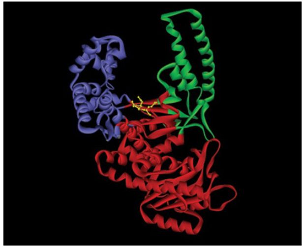
FIGURE 1. The structure of the Klenow fragment from E. coli DNA polymerase I. It has a right hand with fingers (purple), a palm (red), and a thumb (green). The Klenow fragment also includes an exonuclease domain.
Data from: Beese, L. S., et al. 1993. “Structure from Protein Data Bank 1KFD.” Biochemistry 32:14095–14101.
The catalytic reaction in a DNA polymerase occurs at an active site in which a nucleotide triphosphate pairs with an (unpaired) single strand of DNA. The DNA lies across the palm in a groove that is created by the thumb and fingers. FIGURE 2 shows the crystal structure of the Φ T7 enzyme complexed with DNA (in the form of a primer annealed to a template strand) and an incoming nucleotide that is about to be added to the primer. The DNA is in the classic B-form duplex up to the last two base pairs at the 3′ end of the primer, which are in the more open A-form. A sharp turn in the DNA exposes the template base to the incoming nucleotide. The 3′ end of the primer (to which bases are added) is anchored by the fingers and palm. The DNA is held in position by contacts that are made principally with the phosphodiester backbone (thus enabling the polymerase to function with DNA of any sequence).
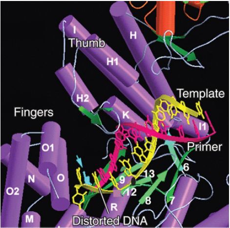
FIGURE 2. The crystal structure of phage T7 DNA polymerase shows that the template strand takes a sharp turn that exposes it to the incoming nucleotide.
Photo courtesy of Charles Richardson and Thomas Ellenberger, Washington University School of Medicine.
In structures of DNA polymerases of this family complexed only with DNA (i.e., lacking the incoming nucleotide), the orientation of the fingers and thumb relative to the palm is more open, with the O helix (O, O1, O2; see Figure 2) rotated away from the palm.
This suggests that an inward rotation of the O helix occurs to grasp the incoming nucleotide and create the active catalytic site. When a nucleotide binds, the fingers domain rotates 60° toward the palm, with the tops of the fingers moving by 30 Å. The thumb domain also rotates toward the palm by 8°. These changes are cyclical: They are reversed when the nucleotide is incorporated into the DNA chain, which then translocates through the enzyme to recreate an empty site.
The exonuclease activity is responsible for removing mispaired bases. The catalytic site of the exonuclease domain is distant from the active site of the catalytic domain, though. The enzyme alternates between polymerizing and editing modes, as determined by a competition between the two active sites for the 3′ primer end of the DNA. Amino acids in the active site contact the incoming base in such a way that the enzyme structure is affected by the structure of a mismatched base. When a mismatched base pair occupies the catalytic site, the fingers cannot rotate toward the palm to bind the incoming nucleotide. This leaves the 3′ end free to bind to the active site in the exonuclease domain, which is accomplished by a rotation of the DNA in the enzyme structure.
 الاكثر قراءة في مواضيع عامة في الاحياء الجزيئي
 اخر الاخبار
اخبار العتبة العباسية المقدسة

الآخبار الصحية


مواضيع ذات صلة
 قسم الشؤون الفكرية يصدر كتاباً يوثق تاريخ السدانة في العتبة العباسية المقدسة
 "المهمة".. إصدار قصصي يوثّق القصص الفائزة في مسابقة فتوى الدفاع المقدسة للقصة القصيرة
 (نوافذ).. إصدار أدبي يوثق القصص الفائزة في مسابقة الإمام العسكري (عليه السلام)


