


النبات

مواضيع عامة في علم النبات
الجذور - السيقان - الأوراق
النباتات الوعائية واللاوعائية
البذور (مغطاة البذور - عاريات البذور)
الطحالب
النباتات الطبية
الحيوان
مواضيع عامة في علم الحيوان
علم التشريح
التنوع الإحيائي
البايلوجيا الخلوية
الأحياء المجهرية
البكتيريا
الفطريات
الطفيليات
الفايروسات
علم الأمراض
الاورام
الامراض الوراثية
الامراض المناعية
الامراض المدارية
اضطرابات الدورة الدموية
مواضيع عامة في علم الامراض
الحشرات
التقانة الإحيائية
مواضيع عامة في التقانة الإحيائية
التقنية الحيوية المكروبية
التقنية الحيوية والميكروبات
الفعاليات الحيوية
وراثة الاحياء المجهرية
تصنيف الاحياء المجهرية
الاحياء المجهرية في الطبيعة
أيض الاجهاد
التقنية الحيوية والبيئة
التقنية الحيوية والطب
التقنية الحيوية والزراعة
التقنية الحيوية والصناعة
التقنية الحيوية والطاقة
البحار والطحالب الصغيرة
عزل البروتين
هندسة الجينات
التقنية الحياتية النانوية
مفاهيم التقنية الحيوية النانوية
التراكيب النانوية والمجاهر المستخدمة في رؤيتها
تصنيع وتخليق المواد النانوية
تطبيقات التقنية النانوية والحيوية النانوية
الرقائق والمتحسسات الحيوية
المصفوفات المجهرية وحاسوب الدنا
اللقاحات
البيئة والتلوث
علم الأجنة
اعضاء التكاثر وتشكل الاعراس
الاخصاب
التشطر
العصيبة وتشكل الجسيدات
تشكل اللواحق الجنينية
تكون المعيدة وظهور الطبقات الجنينية
مقدمة لعلم الاجنة
الأحياء الجزيئي
مواضيع عامة في الاحياء الجزيئي
علم وظائف الأعضاء
الغدد
مواضيع عامة في الغدد
الغدد الصم و هرموناتها
الجسم تحت السريري
الغدة النخامية
الغدة الكظرية
الغدة التناسلية
الغدة الدرقية والجار الدرقية
الغدة البنكرياسية
الغدة الصنوبرية
مواضيع عامة في علم وظائف الاعضاء
الخلية الحيوانية
الجهاز العصبي
أعضاء الحس
الجهاز العضلي
السوائل الجسمية
الجهاز الدوري والليمف
الجهاز التنفسي
الجهاز الهضمي
الجهاز البولي
المضادات الميكروبية
مواضيع عامة في المضادات الميكروبية
مضادات البكتيريا
مضادات الفطريات
مضادات الطفيليات
مضادات الفايروسات
علم الخلية
الوراثة
الأحياء العامة
المناعة
التحليلات المرضية
الكيمياء الحيوية
مواضيع متنوعة أخرى
الانزيمات

Nucleases
 المؤلف:
JOCELYN E. KREBS, ELLIOTT S. GOLDSTEIN and STEPHEN T. KILPATRICK
المؤلف:
JOCELYN E. KREBS, ELLIOTT S. GOLDSTEIN and STEPHEN T. KILPATRICK
 المصدر:
LEWIN’S GENES XII
المصدر:
LEWIN’S GENES XII
 الجزء والصفحة:
 5-3-2021
 3353
3353


+
-
20
Nucleases
KEY CONCEPTS
-Nucleases hydrolyze an ester bond within a phosphodiester bond.
-Phosphatases hydrolyze the ester bond in a phosphomonoester bond.
-Nucleases have a multiplicity of specificities.
-Restriction endonucleases cleave DNA into defined fragments.
-A map can be generated by using the overlaps between the fragments generated by different restriction enzymes.
Nucleases are one of the most valuable tools in a molecular biology laboratory. One class of enzymes, the restriction endonucleases (discussed shortly), was critical for the cloning revolution. Nucleases are enzymes that degrade nucleic acids, the opposite function of polymerases. They hydrolyze, or break, an ester bond in a phosphodiester linkage between adjacent nucleotides in a polynucleotide chain, as shown in FIGURE1.
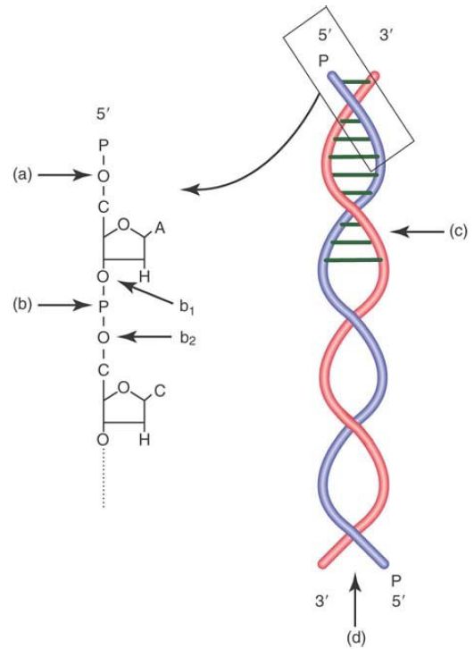
FIGURE 1. The target of a phosphatase is shown in (a), a terminal phosphomonoester bond. The target of a nuclease is shown in (b), the phosphodiester bond between two adjacent nucleotides. Note that the nuclease can cleave either the first ester bond from the 3′ end of the terminal nucleotide (b1 ) or the second ester bond from the 5′ end of the next nucleotide (b2 ). Nucleases can cleave internal bonds (c) as an endonuclease, or begin at an end and progress into the fragment (d) as an exonuclease.
There is another, related class of enzymes that can hydrolyze an ester bond in a nucleotide chain—a monoesterase, usually called a phosphatase. The critical difference between a phosphatase and a nuclease is shown in Figure 1. A phosphatase can only hydrolyze a terminal ester bond linking a phosphate (or di- or triphosphate) to a terminal nucleotide at the 3′ or 5′ end, whereas a nuclease can hydrolyze an internal ester bond in a diester link, between adjacent bases.
Phosphatases are important enzymes in the laboratory because they allow the removal of a terminal phosphate from a polynucleotide chain. This is often required for a subsequent step of connecting, or ligating, chains together. This also allows one to replace the phosphate with a radioactive P molecule.
Nucleases can be divided into groups based on a number of different features. We can distinguish between endonucleases and exonucleases as shown in Figure 1. An endonuclease can hydrolyze internal bonds within a polynucleotide chain, whereas an exonuclease must begin at the end of a chain and hydrolyze from that end position.
The specificity of nucleases ranges from none to extreme. Nucleases can be specific for DNA, as DNases, or RNA, as RNases, or even be specific for a DNA/RNA hybrid, as RNaseH (which cleaves the RNA strand of a hybrid duplex). Nucleases can be specific for either single-stranded nucleotide chains, duplex chains, or both.
When a nuclease—either endo- or exo-—hydrolyzes an ester bond in a phosphodiester linkage, it will have specificity for either of the two ester bonds, generating either 5′ nucleotides or 3′ nucleotides, as shown in Figure 2.1. An exonuclease can attack a polynucleotide chain from either the 5′ end and hydrolyze 5′ to 3′ or attack from the 3′ end and hydrolyze 3′ to 5′ (Figure 1).
Nucleases might have a sequence preference, such as pancreatic RNase A, which preferentially cuts after a pyrimidine, or T1 RNase, which cuts single-stranded RNA chains after a G. At the extreme end of sequence specificity lie the restriction endonucleases, usually called restriction enzymes. These are endonucleases from eubacteria and Archaea that recognize a specific DNA
sequence. Their name typically derives from the bacteria in which they were discovered. For example, EcoR1 is the first restriction enzyme from an Escherichia coli R strain.
Broadly speaking, there are three different classes of restriction enzymes and several subclasses. In 1978, the Nobel Prize in Medicine was awarded to Daniel Nathans, Werner Arber, and Hamilton Smith for the discovery of restriction endonucleases. It was this discovery that enabled scientists to develop the methods to clone DNA, as shown in the next section. Thousands of restriction enzymes are known, many of which are now commercially available. Restriction enzymes have to do two things: (1) recognize a specific sequence, and (2) cut, or restrict, at or near that sequence.
The type II restriction enzymes (with several subgroups) are the most common. Type II enzymes are distinguished because the recognition site and cleavage site are the same. These sites range in length from 4 to 8 base pairs (bp). The sites are typically inversely palindromic, that is, reading the same forward and backward on complementary strands, as shown in FIGURE 2.
Restriction enzymes can cut the DNA in two different ways, as demonstrated in Figure .2. The first and more common is a staggered cut, which leaves single-stranded overhangs, or “sticky ends.” The overhang can be a 3′ or a 5′ overhang. The second way is a blunt double-stranded cut, which does not leave an overhang.
An additional level of specificity determines whether the enzyme will cut DNA containing a methylated base. The degree of specificity in the site also varies. Most enzymes are very specific, whereas some will allow multiple bases at one or two positions within the site.
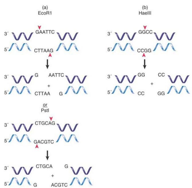
FIGURE 2. (a) A restriction endonuclease may cleave its recognition site and make a staggered cut, leaving a 5′ overhang or a 3′ overhang. (b) A restriction endonuclease may cleave its recognition site and make a blunt end cut.
Restriction enzymes from different bacteria can have the same recognition site but cut the DNA differently. One might make a blunt cut and the other might make a staggered cut, or one might leave a 3′ overhang, whereas the second might leave a 5′ overhang. These different enzymes are called isoschizomers.
Types I and III enzymes differ from type II enzymes in that the recognition site and cleavage site are different and are usually not palindromes. With a type I enzyme, the cleavage site can be up to 1,000 bp away from the recognition site. Type III enzymes have closer cleavage sites, usually 20 to 30 bp away.
A restriction map represents a linear sequence of the sites at which particular restriction enzymes find their targets. When a DNA molecule is cut with a suitable restriction enzyme, it is cleaved into distinct, negatively charged fragments. These fragments can be separated on the basis of their size by gel electrophoresis (described later, in the section DNA Separation Techniques). By analyzing the restriction fragments of DNA, it is possible to generate a map of the original molecule in the form shown in FIGURE 3. The map shows the positions at which particular restriction enzymes cut DNA. The DNA is divided into a series of regions of defined lengths that lie between sites recognized by the restriction enzymes. A restriction map can be obtained for any sequence of DNA, irrespective of whether we have any knowledge of its function. If the sequence of the DNA is known, we can generate a restriction map in silico by simply searching for the recognition sites of known enzymes. Knowing the restriction map of a DNA sequence of interest is extremely valuable in DNA cloning, which is described in the next section.
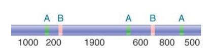
FIGURE 3 A restriction map is a linear sequence of sites separated by defined distances on DNA. The map identifies the three sites cleaved by enzyme A and the two sites cleaved by enzyme B. Thus, A produces four fragments, which overlap those of B, and B produces three fragments, which overlap those of A.
 الاكثر قراءة في مواضيع عامة في الاحياء الجزيئي
 اخر الاخبار
اخبار العتبة العباسية المقدسة

الآخبار الصحية


مواضيع ذات صلة
 قسم الشؤون الفكرية يصدر كتاباً يوثق تاريخ السدانة في العتبة العباسية المقدسة
 "المهمة".. إصدار قصصي يوثّق القصص الفائزة في مسابقة فتوى الدفاع المقدسة للقصة القصيرة
 (نوافذ).. إصدار أدبي يوثق القصص الفائزة في مسابقة الإمام العسكري (عليه السلام)


