


النبات

مواضيع عامة في علم النبات
الجذور - السيقان - الأوراق
النباتات الوعائية واللاوعائية
البذور (مغطاة البذور - عاريات البذور)
الطحالب
النباتات الطبية
الحيوان
مواضيع عامة في علم الحيوان
علم التشريح
التنوع الإحيائي
البايلوجيا الخلوية
الأحياء المجهرية
البكتيريا
الفطريات
الطفيليات
الفايروسات
علم الأمراض
الاورام
الامراض الوراثية
الامراض المناعية
الامراض المدارية
اضطرابات الدورة الدموية
مواضيع عامة في علم الامراض
الحشرات
التقانة الإحيائية
مواضيع عامة في التقانة الإحيائية
التقنية الحيوية المكروبية
التقنية الحيوية والميكروبات
الفعاليات الحيوية
وراثة الاحياء المجهرية
تصنيف الاحياء المجهرية
الاحياء المجهرية في الطبيعة
أيض الاجهاد
التقنية الحيوية والبيئة
التقنية الحيوية والطب
التقنية الحيوية والزراعة
التقنية الحيوية والصناعة
التقنية الحيوية والطاقة
البحار والطحالب الصغيرة
عزل البروتين
هندسة الجينات
التقنية الحياتية النانوية
مفاهيم التقنية الحيوية النانوية
التراكيب النانوية والمجاهر المستخدمة في رؤيتها
تصنيع وتخليق المواد النانوية
تطبيقات التقنية النانوية والحيوية النانوية
الرقائق والمتحسسات الحيوية
المصفوفات المجهرية وحاسوب الدنا
اللقاحات
البيئة والتلوث
علم الأجنة
اعضاء التكاثر وتشكل الاعراس
الاخصاب
التشطر
العصيبة وتشكل الجسيدات
تشكل اللواحق الجنينية
تكون المعيدة وظهور الطبقات الجنينية
مقدمة لعلم الاجنة
الأحياء الجزيئي
مواضيع عامة في الاحياء الجزيئي
علم وظائف الأعضاء
الغدد
مواضيع عامة في الغدد
الغدد الصم و هرموناتها
الجسم تحت السريري
الغدة النخامية
الغدة الكظرية
الغدة التناسلية
الغدة الدرقية والجار الدرقية
الغدة البنكرياسية
الغدة الصنوبرية
مواضيع عامة في علم وظائف الاعضاء
الخلية الحيوانية
الجهاز العصبي
أعضاء الحس
الجهاز العضلي
السوائل الجسمية
الجهاز الدوري والليمف
الجهاز التنفسي
الجهاز الهضمي
الجهاز البولي
المضادات الميكروبية
مواضيع عامة في المضادات الميكروبية
مضادات البكتيريا
مضادات الفطريات
مضادات الطفيليات
مضادات الفايروسات
علم الخلية
الوراثة
الأحياء العامة
المناعة
التحليلات المرضية
الكيمياء الحيوية
مواضيع متنوعة أخرى
الانزيمات

DNA Is the Genetic Material of Bacteria and Viruses
 المؤلف:
JOCELYN E. KREBS, ELLIOTT S. GOLDSTEIN and STEPHEN T. KILPATRICK
المؤلف:
JOCELYN E. KREBS, ELLIOTT S. GOLDSTEIN and STEPHEN T. KILPATRICK
 المصدر:
LEWIN’S GENES XII
المصدر:
LEWIN’S GENES XII
 الجزء والصفحة:
 23-2-2021
 3360
3360


+
-
20
DNA Is the Genetic Material of Bacteria and Viruses
KEY CONCEPTS
Bacterial transformation provided the first evidence that DNA is the genetic material of bacteria. We can transfer genetic properties from one bacterial strain to another by extracting DNA from the first strain and adding it to the second strain.
Phage infection showed that DNA is the genetic material of some viruses. When the DNA and protein components of bacteriophages are labeled with different radioactive isotopes, only the DNA is transmitted to the progeny phages produced by infecting bacteria.
The idea that the genetic material of organisms is DNA has its roots in the discovery of transformation by Frederick Griffith in 1928. The bacterium Streptococcus (formerly Pneumococcus) pneumoniae kills mice by causing pneumonia. The virulence of the bacterium is determined by its capsular polysaccharide, which allows the bacterium to escape destruction by its host. Several types of S. pneumoniae have different capsular polysaccharides, but they all have a smooth “S” appearance. Each of the S types can give rise to variants that fail to produce the capsular polysaccharide and therefore have a rough “R” surface (consisting of the material that was beneath the capsular polysaccharide). The R types are avirulent and do not kill the mice, because the absence of the polysaccharide capsule allows the animal’s immune system
to destroy the bacteria.
When S bacteria are killed by heat treatment, they can no longer harm the animal. FIGURE 1, however, shows that when heatkilled S bacteria and avirulent R bacteria are jointly injected into a mouse, it dies as the result of a pneumonia infection. Virulent S bacteria can be recovered from the mouse’s blood.
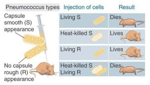
FIGURE 1. Neither heat-killed S-type nor live R-type bacteria can kill mice, but simultaneous injection of both can kill mice just as effectively as the live S type.
In this experiment, the heat-killed S bacteria were of type III and the live R bacteria had been derived from type II. The virulent bacteria recovered from the mixed infection had the smooth coat of type III. So, some property of the dead IIIS bacteria can transform the live IIR bacteria so that they make the capsular polysaccharide and become virulent. FIGURE 2 shows the identification of the component of the dead bacteria responsible for transformation.
This was called the transforming principle. It was purified in a cell-free system in which extracts from the dead IIIS bacteria were added to the live IIR bacteria before being plated on agar and assayed for transformation (FIGURE 3). Purification of the transforming principle in 1944 by Avery, MacLeod, and McCarty showed that it is DNA.
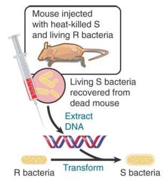
FIGURE 2 The DNA of S-type bacteria can transform R-type bacteria into the same S type.
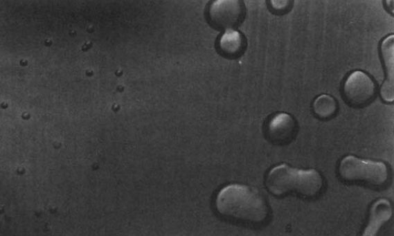
FIGURE 3. Rough (left) and smooth (right) colonies of S. pneumoniae.
Having shown that DNA is the genetic material of bacteria, the next step was to demonstrate that DNA is the genetic material in a quite different system. Phage T2 is a virus that infects the bacterium Escherichia coli. When phage particles are added to bacteria, they attach to the outside surface, some material enters the cell, and then approximately 20 minutes later each cell bursts open, or lyses, to release a large number of progeny phage.
FIGURE 4 illustrates the results of an experiment conducted in 1952 by Alfred Hershey and Martha Chase in which bacteria were infected with T2 phages that had been radioactively labeled either in their DNA component (with phosphorus-32 [32P]) or in their protein component (with sulfur-35 [35S]). The infected bacteria were agitated in a blender and two fractions were separated by centrifugation. One fraction, containing the empty phage “ghosts” that were released from the surface of the bacteria, consisted of protein and contained approximately 80% of the S label. The other fraction consisted of the infected bacteria themselves and contained approximately 70% of the P label. Previously, it had been shown that phage replication occurs intracellularly so that the genetic material of the phage would have to enter the cell during infection.
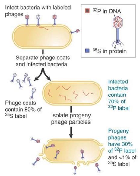
FIGURE 4. The genetic material of phage T2 is DNA.
Most of the P label was present in the fraction containing infected bacteria. The progeny phage particles produced by the infection contained approximately 30% of the original P label. The progeny received less than 1% of the protein contained in the original phage population. This experiment directly showed that only the DNA of the parent phages enters the bacteria and becomes part of the progeny phages, which is exactly the expected behavior of genetic material.
The phage possesses genetic material with properties analogous to those of cellular genomes: Its traits are faithfully expressed and are subject to the same rules that govern inheritance of cellular traits. The case of T2 reinforces the general conclusion that DNA is the genetic material of the genome of a cell or a virus.
 الاكثر قراءة في مواضيع عامة في الاحياء الجزيئي
 اخر الاخبار
اخبار العتبة العباسية المقدسة

الآخبار الصحية


مواضيع ذات صلة
 قسم الشؤون الفكرية يصدر كتاباً يوثق تاريخ السدانة في العتبة العباسية المقدسة
 "المهمة".. إصدار قصصي يوثّق القصص الفائزة في مسابقة فتوى الدفاع المقدسة للقصة القصيرة
 (نوافذ).. إصدار أدبي يوثق القصص الفائزة في مسابقة الإمام العسكري (عليه السلام)


