![]()


النبات

مواضيع عامة في علم النبات
الجذور - السيقان - الأوراق
النباتات الوعائية واللاوعائية
البذور (مغطاة البذور - عاريات البذور)
الطحالب
النباتات الطبية
الحيوان
مواضيع عامة في علم الحيوان
علم التشريح
التنوع الإحيائي
البايلوجيا الخلوية
الأحياء المجهرية
البكتيريا
الفطريات
الطفيليات
الفايروسات
علم الأمراض
الاورام
الامراض الوراثية
الامراض المناعية
الامراض المدارية
اضطرابات الدورة الدموية
مواضيع عامة في علم الامراض
الحشرات
التقانة الإحيائية
مواضيع عامة في التقانة الإحيائية
التقنية الحيوية المكروبية
التقنية الحيوية والميكروبات
الفعاليات الحيوية
وراثة الاحياء المجهرية
تصنيف الاحياء المجهرية
الاحياء المجهرية في الطبيعة
أيض الاجهاد
التقنية الحيوية والبيئة
التقنية الحيوية والطب
التقنية الحيوية والزراعة
التقنية الحيوية والصناعة
التقنية الحيوية والطاقة
البحار والطحالب الصغيرة
عزل البروتين
هندسة الجينات
التقنية الحياتية النانوية
مفاهيم التقنية الحيوية النانوية
التراكيب النانوية والمجاهر المستخدمة في رؤيتها
تصنيع وتخليق المواد النانوية
تطبيقات التقنية النانوية والحيوية النانوية
الرقائق والمتحسسات الحيوية
المصفوفات المجهرية وحاسوب الدنا
اللقاحات
البيئة والتلوث
علم الأجنة
اعضاء التكاثر وتشكل الاعراس
الاخصاب
التشطر
العصيبة وتشكل الجسيدات
تشكل اللواحق الجنينية
تكون المعيدة وظهور الطبقات الجنينية
مقدمة لعلم الاجنة
الأحياء الجزيئي
مواضيع عامة في الاحياء الجزيئي
علم وظائف الأعضاء
الغدد
مواضيع عامة في الغدد
الغدد الصم و هرموناتها
الجسم تحت السريري
الغدة النخامية
الغدة الكظرية
الغدة التناسلية
الغدة الدرقية والجار الدرقية
الغدة البنكرياسية
الغدة الصنوبرية
مواضيع عامة في علم وظائف الاعضاء
الخلية الحيوانية
الجهاز العصبي
أعضاء الحس
الجهاز العضلي
السوائل الجسمية
الجهاز الدوري والليمف
الجهاز التنفسي
الجهاز الهضمي
الجهاز البولي
المضادات الحيوية
مواضيع عامة في المضادات الحيوية
مضادات البكتيريا
مضادات الفطريات
مضادات الطفيليات
مضادات الفايروسات
علم الخلية
الوراثة
الأحياء العامة
المناعة
التحليلات المرضية
الكيمياء الحيوية
مواضيع متنوعة أخرى
الانزيمات

Glycogen Metabolism
 المؤلف:
Denise R. Ferrier
المؤلف:
Denise R. Ferrier
 المصدر:
Lippincott Illustrated Reviews: Biochemistry
المصدر:
Lippincott Illustrated Reviews: Biochemistry
 الجزء والصفحة:
 26-9-2021
 1340
1340


Glycogen Metabolism
The main stores of glycogen in the body are found in skeletal muscle, where they serve as a fuel reserve for the synthesis of ATP during muscle contraction, and in the liver, where they are used to maintain the blood glucose concentration, particularly during the early stages of a fast.
Glycogen is a highly branched polymer of α-D-glucose. The primary glycosidic bond is an α(1→4) linkage. After about 8–14 glucosyl residues, there is a branch containing an α(1→6) linkage. Uridine diphosphate (UDP)-glucose, the building block of glycogen, is synthesized from glucose 1-phosphate and UTP by UDP–glucose pyrophosphorylase (Fig. 1).
Glucose from UDP-glucose is transferred to the nonreducing ends of glycogen chains by primer-requiring glycogen synthase, which makes the α(1→4) linkages. The primer is made by glycogenin. Branches are formed by amylo-α(1→4)→α(1→6)-transglycosylase (a 4:6 transferase), which transfers a set of six to eight glucosyl residues from the nonreducing end of the glycogen chain (breaking an α(1→4) linkage), and making an α(1→6) linkage to another residue in the chain. Pyridoxal phosphate–requiring glycogen phosphorylase cleaves the α(1→4) bonds between glucosyl residues at the nonreducing ends of the glycogen chains, producing glucose 1-phosphate. This sequential degradation continues until four glucosyl units remain before a branch point. The resulting structure is called a limit dextrin that is degraded by the bifunctional debranching enzyme. Oligo-α(1→4)→α(1→4)-glucantransferase (a 4:4 transferase) activity removes the outer three of the four glucosyl residues at a branch and transfers them to the nonreducing end of another chain, where they can be released as glucose 1-phosphate by glycogen phosphorylase. The remaining single glucose residue attached in an α(1→6) linkage is removed hydrolytically by the amylo-α(1→6) glucosidase activity of debranching enzyme, releasing free glucose.
Glucose 1-phosphate is converted to glucose 6-phosphate by phosphoglucomutase. In muscle, glucose 6-phosphate enters glycolysis. In liver, the phosphate is removed by glucose 6-phosphatase (an enzyme of the endoplasmic reticular membrane), releasing free glucose that can be used to maintain blood glucose levels at the beginning of a fast. A deficiency of the phosphatase causes glycogen storage disease Ia (von Gierke disease) and results in an inability of the liver to provide free glucose to the body during a fast. It affects both glycogen degradation and gluconeogenesis. Glycogen synthesis and degradation are reciprocally regulated to meet whole-body needs by the same hormonal signals (namely, an elevated insulin level results in overall increased glycogenesis and decreased glycogenolysis, whereas an elevated glucagon, or epinephrine, level causes the opposite effects). Key enzymes are phosphorylated by a family of protein kinases, some of which are dependent on cyclic adenosine monophosphate (cAMP), a compound increased by glucagon and epinephrine. Phosphate groups are removed by protein phosphatase-1 (active when its inhibitor is inactive in response to elevated insulin levels). In addition to this covalent regulation, glycogen synthase, phosphorylase kinase, and phosphorylase are allosterically regulated to meet tissues’ needs. In the well-fed state, glycogen synthase is activated by glucose 6-phosphate, but glycogen phosphorylase is inhibited by glucose 6-phosphate as well as by ATP. In the liver, free glucose also serves as an allosteric inhibitor of glycogen phosphorylase. The rise in calcium in muscle during exercise and in liver in response to epinephrine activates phosphorylase kinase by binding to the enzyme’s calmodulin subunit. This allows the enzyme to activate glycogen phosphorylase, thereby causing glycogen degradation. AMP activates glycogen phosphorylase (myophosphorylase) in muscle.
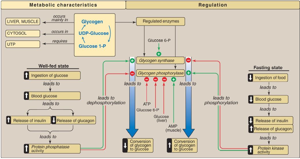 Figure 1: Key concept map for glycogen metabolism in the liver. [Note: Glycogen phosphorylase is phosphorylated by phosphorylase kinase, the “b” form of which can be activated by calcium.] UDP and UTP = uridine di- and triphosphates; P = phosphate; AMP = adenosine monophosphate.


 قسم الشؤون الفكرية يصدر مجموعة قصصية بعنوان (قلوب بلا مأوى)
 قسم الشؤون الفكرية يصدر مجموعة قصصية بعنوان (قلوب بلا مأوى)
 قسم الشؤون الفكرية يصدر كتاب (سر الرضا) ضمن سلسلة (نمط الحياة)


