آخر المواضيع المضافة

 النبات
الحيوان
الأحياء المجهرية
علم الأمراض
التقانة الإحيائية
التقنية الحيوية المكروبية
التقنية الحياتية النانوية
علم الأجنة
الأحياء الجزيئي
علم وظائف الأعضاء
الغدد
المضادات الحيوية
النبات
الحيوان
الأحياء المجهرية
علم الأمراض
التقانة الإحيائية
التقنية الحيوية المكروبية
التقنية الحياتية النانوية
علم الأجنة
الأحياء الجزيئي
علم وظائف الأعضاء
الغدد
المضادات الحيوية| Exocytosis |


|
|
Read More
Date: 1-12-2015
Date: 10-12-2020
Date: 2-6-2021
|
Exocytosis
Exocytosis occurs in eukaryotic cells when the membrane of a cytoplasmic vesicle fuses with the plasma membrane, exposing the contents of the vesicle and the lumenal layer of the phospholipid bilayer to the outside world.
1. Examples of Exocytosis
Most cells can secrete proteins. In bacteria, the proteins can be unfolded and then threaded, or translocated, across the plasma membrane. Eukaryotes translocate newly synthesized proteins into an intracellular organelle, the endoplasmic reticulum. Proteins travel from the endoplasmic reticulum by way of the Golgi complex, to the plasma membrane. Transport is effected by packaging the newly synthesized proteins in secretory vesicles, which must fuse their membrane with that of the plasma membrane to release their contents to the extracellular space. Thus eukaryotic cells that secrete proteins must have the molecular machinery to allow exocytosis at the cell surface. Exocytosis must be highly efficient, since few secretory vesicles are seen in most cells.
Sometimes newly synthesized proteins passing through the Golgi complex are diverted selectively into cytoplasmic storage vesicles, usually called dense core secretory granules (1). To allow cytoplasmic storage, exocytosis of such secretory granules is inhibited, causing the accumulation of granules in the cytoplasm or along the plasma membrane. Accumulation of storage or secretory granules usually gives cells a characteristic morphology; examples are the granulocytes of the hematopoietic system, endocrine and exocrine cells, prefertilization eggs filled with cortical granules, and protozoa such as Paramecium and Tetrahymena . A signal to the outside of such protein-storage cells can remove the inhibition to exocytosis and trigger a massive efflux of protein, a process referred to as regulated secretion.
Some endocrine cells, such as chromaffin cells or neurons, actively transport small molecules from the cytoplasm into the lumen of the dense core secretory granules, giving them high internal concentrations of both protein and the small molecules (2). The energy for the active transport comes from a proton gradient across the secretory granule membrane (3). This property is also found in bone marrow-derived cells. Mast cells, for example, package histamine to high concentrations inside their secretory granules. When cells of this type undergo exocytosis, along with the stored proteins they release massive quantities of the small molecules, which usually are identical, or are related to, neurotransmitters.
In addition to having a biosynthetic pathway for protein secretion, many eukaryotic cells have an endocytosis pathway that internalizes plasma membrane components and returns most of them to the cell surface (4). This membrane recycling pathway allows a cell to move membrane to a site where it might be more needed; for example, a region of cell growth or migration (5). The recycling pathway also allows a cell to internalize nutrients bound to receptors, to remove the nutrients, and to send the receptors back to the cell surface for more. In the final step, a membrane vesicle on the recycling pathway must fuse with the plasma membrane (that is, it must undergo exocytosis). Thus, the exocytosis machinery is essential for endocytotic recycling, as well as for protein secretion (Fig. 1) .
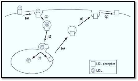
Figure 1. Example of exocytosis in endocytosis. Exocytosis is used in other processes besides delivery of secretory proteins to the cell surface. Here, using the trafficking of low-density lipoprotein (LDL) receptor as an example, we see how exocytosis is an integral part of endocytic trafficking. (a) Cholesterol-containing LDLs bind to their receptors and (b) are internalized by endocytosis. (c) The endocytic vesicles fuse with an endosome (d) where LDL dissociates from its receptor. While LDL is eventually broken down for utilization of the cholesterol (ef), the LDL receptor is recycled back to the cell surface by exocytosis (g) so that it can be reused.
As with protein secretion, there appear to be constitutive and regulated exocytotic steps associated with membrane recycling. Membrane vesicles carrying nutrient receptors, such as the receptors for transferrin and low-density lipoprotein, fuse rapidly with the plasma membrane, independently of an external signal. In specialized cases, exocytosis is inhibited, causing an accumulation of endocytotically derived membrane vesicles (6). Two examples of such storage vesicles are synaptic vesicles and vesicles storing intracellularly a special type of glucose transporter, GLUT4 (glucose transporter 4) (7). Regulated exocytosis of the endocytotically derived vesicles can occur when the inhibition is removed, by an electrical signal in the case of the nerve cell, and by insulin in the case of GLUT4-storing fat and muscle cells.
Fortunately, similar molecular machinery seems to be used by all forms of exocytosis, regulated or constitutive, biosynthetic, or plasma-membrane recycling. Furthermore, the molecules that regulate exocytosis appear to be conserved, as are the transporters that put neurotransmitters into dense core secretory granules and into synaptic vesicles.
2. Measurements of Exocytosis
A common but crude measure of exocytosis is the appearance in the extracellular medium of the contents of a secretory vesicle that has undergone exocytosis. Thus exocytosis of synaptic vesicles (see Secretory Vesicles/Granules) at the nerve terminal causes release of neurotransmitter, and exocytosis of secretory granules from b-cells of the pancreatic islets of Langerhans releases insulin. Extracellular neurotransmitters and hormones can be measured biochemically, as can the constitutive secretion of enzymes such as invertase by yeast or proteinases by mammalian cells. Biochemical measurements are usually incapable of measuring the kinetics of exocytosis with accuracy.
The release of neurotransmitters, however, can be measured electrophysiologically with much greater precision. Most neurotransmitters activate the postsynaptic target cell by binding to closed ion channels and causing them to open. Measurement of current flow through the postsynaptic ion channels gives an almost instantaneous assay of the exocytosis rates in the presynaptic nerve terminal. The precision and sensitivity of electrophysiological measurements, pioneered by Dr. Bernhard Katz and his coworkers at University College, London, allowed detection of a quantum of neurotransmitter release, lasting a millisecond or so, generated by exocytosis of the contents of a single synaptic vesicle. Electrophysiology revealed that exocytosis is very rapid, occurring within 100 µs of a stimulus to the nerve terminal, and that the concentration of the neurotransmitter in the synaptic vesicle is very high, greater than 100 mM.
Postsynaptic currents generated by presynaptic release of neurotransmitter accurately measured the exocytosis because the receptor channels were 10 to 20 nm from the exocytosis site. Release of biogenic amines from cells such as chromaffin cells can likewise be measured rapidly and quantitatively by placing a carbon electrode close to the cell and detecting the biogenic amine by its redox potential (8). This technique was first applied to exocytosis by Wightman's group and is called amperometry.
Another technique widely used to study exocytosis uses the cell-surface capacitance. The electrical capacitance across the plasma membrane of a cell is proportional to its area. When exocytosis occurs, the capacitance increases because of the addition of membrane (9, 10). The sensitivity of capacitance measurements is such that it can measure the addition of the membrane of a single secretory granule. It cannot yet detect the addition of a single synaptic vesicle, since they are too small (50 nm diameter). When the capacitance signal is big enough, however, the capacitance changes give an instantaneous measurement of exocytosis.
To measure capacitance, a cell must be attached to a “patch electrode,” which allows electrical communication between the salt solution inside the electrode and the cytoplasm of the cell.
Video techniques for quantifying fluorescent markers in cells and recording changes in fluorescence intensity as a function of time are becoming increasingly sophisticated. Video-microscopy techniques are used to study exocytosis by loading synaptic vesicles with a membrane-impermeable fluorescent dye (11, 12). Each synaptic vesicle can contain about 30 dye molecules. When the membrane of a synaptic vesicle fuses with that of the plasma membrane, the intensity of the fluorescence decreases by a fixed amount, which can be measured using sensitive detection devices. The dye technique for studying exocytosis was pioneered by William Betz and his colleagues at the University of Colorado.
When the secretory vesicle is big enough, it is possible to observe exocytosis in the light microscope. Some protozoa, such as Paramecium or Tetrahymena, have large cytoplasmic vesicles, packed with protein, that are docked at the plasma membrane. When the light microscope is used (13), the dense contents of the secretory granules can be seen to disappear when exocytosis is triggered. The convenience of a visual assay for exocytosis has made it possible to screen for secretion mutants in organisms of this type. Exocytosis of secretory granule content can also be detected visually in sea urchin eggs and in some classes of mast cell.
3. Mechanism of Exocytosis
Exocytosis takes place in several stages. First the membrane of the vesicle recognizes the plasma membrane and forms a close association with it. Then the phospholipid bilayers are rearranged so that the membranes of the vesicle and those of the plasma membrane become continuous. Finally, the curved vesicle membrane flattens out, adopting the curvature of the plasma membrane.
Association of the cytoplasmic vesicle with the membrane is sometimes called docking . There are two types of vesicle docking, which sometimes get confused. Since exocytosis is not a random event but requires docking specifically between a vesicle and plasma membrane, the two membranes must recognize each other, presumably through protein–protein interactions. The recognition and fusion steps must be rapid processes in most cells, because few vesicles are seen adjacent to the plasma membrane or in the process of fusion. In contrast, in regulated secretory cells, such as neurons and endocrine cells, the secretory and synaptic vesicles are in a stable association with the plasma membrane. The long-lasting docking in neurons and neuroendocrine cells is presumably an adaptation that allows very rapid exocytosis in response to an extracellular signal. To avoid confusing the two forms of docking, we discuss first the generic, short-term docking, present in all eukaryotic cells.
Many of the molecular insights into exocytosis derive from mutations that affect protein secretion in yeast. Such mutants can have defects in all stages of the protein export pathway. Particularly relevant to exocytosis are the mutations that accumulate post-Golgi secretory vesicles in their cytoplasm. Of the three sets of gene products that affect exocytosis, the ones that are understood in most detail are the SNARE proteins (14). The name SNARE (SNAp REceptor( derives from one of the ways the proteins were discovered (discussed later) and is useful to describe their function. A v-SNARE protein on a vesicle targeted for exocytosis interacts with a t-SNARE on the target plasma membrane. The first identified member of the v-SNARE family was the synaptic vesicle protein initially called VAMP (Veside Associated Membrane Protein) (15) and later synaptobrevin (16). The characteristics of v-SNAREs on secretory vesicles include (1) a vestigial lumenal or extracytoplasmic domain at the C-terminus, which can be as small as two amino acid residues; (2) a single conserved transmembrane domain; (3) a highly conserved juxtamembrane region that has the predicted sequence of a coiled coil, and (4) a variable N-terminal domain. At the cell surface, the v-SNARE is believed to interact with two t-SNAREs. One is a member of the syntaxin family (17), which also has a small lumenal C-terminal domain, a single transmembrane domain, and a coiled-coil region. The third, a member of the SNAP (synaptosomal-associated protein)-25 family (18), is a cytoplasmic peripheral protein, also with a coiled-coil domain, and is held to the plasma membrane by covalently attached palmitoyl groups. The three proteins are believed to form a ternary complex via their helical domains, which facilitates fusion (19). All well-studied exocytotic events, from yeast to mammalian cells, also appear to involve close homologues of these three SNARE proteins (20). Even synaptic-vesicle exocytosis, where the fusing vesicle is endocytotically derived, uses SNARE proteins. This has been shown using mutations in Drosophila that affect the neuronal SNAREs VAMP/synaptobrevin or syntaxin (21, 22). The involvement of SNAREs can also be verified by use of bacterial neurotoxins. Tetanus toxin and the botulinum neurotoxins A to G are endopeptidases that specifically hydrolyze the SNARE molecules (23. ( Exocytosis from nerve terminals and endocrine cells is blocked by these neurotoxins, providing evidence for the involvement of the SNARE complex even in regulated exocytosis (24).
SNARE complexes form readily, even with purified components in the presence of detergents. Given their high proclivity for spontaneous interaction, it is no surprise that the SNAREs appear to be sequestered in complexes that limit their reactivity. The v-SNAREs in synaptic vesicles )VAMP/synaptobrevin) is sequestered by the synaptic-vesicle protein synaptophysin, which has four transmembrane segments (25). The corresponding t-SNARE, syntaxin, is strongly bound to a cytosolic protein, which is called the sec1 gene product in yeast and nsec1, for neuronal sec1, in the nervous system (26, 27). Since SNARE complexes are so energetically stable, energy must be expended to separate the SNAREs. Separation of the complex involves the ATPase NSF (N-ethyl maleimide sensitive factor) and two accessory proteins, called a- and b-SNAP (28). For NSF to bind, the SNAPs must bind the SNARE complexes. Thus the SNARE complexes received their name, SNAP receptors. The acronym SNAP stands for soluble NSF-acceptor protein in this case. For the t-SNARE SNAP-25, the acronym denotes synaptosomal-associated protein. The identity of the acronyms is coincidental but confusing. Currently, it is thought that NSF might act to dissociate the SNARE complex before fusion, but this is controversial (29, 30) (Fig. 2).
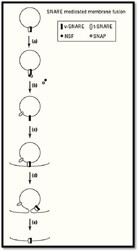
Figure 2. SNARE-mediated membrane fusion. (a) NSF and SNAP proteins bind to v-SNARE/t-SNARE complexes on membranes. (b) ATP hydrolysis by NSF causes dissociation of the SNARE complex so that (c) the v-SNARE can form a complex with a t-SNARE on a target membrane. (d) By an unknown mechanism complexes formed with SNAREs on opposing membranes are thought to promote membrane fusion (e), ending with the SNARE complex on the same membrane. NSF/SNAP must dissociate the complex again before either of the SNAREs can be reused.
Exocytosis in yeast is also inhibited by a mutation in a small, ras-like, GTPase, the sec4 gene product (31) . The sec4 protein is a member of a large class of small GTPases, the rab proteins. All secretory vesicles have a rab protein. In the case of synaptic vesicles, it is rab3A. Rab proteins appear to interact with a complex of proteins, of which the rabaptin complex is the prototype (32). The rab system seems to parallel the SNARE system in docking secretory vesicles at the plasma membrane.
Thus, yeast mutations in the rab pathway can be rescued by overexpression of genes in the SNARE pathway (24). Another protein complex that appears to determine the site of exocytosis in yeast and mammalian cells is the exocyst (33, 34. (
The final class of players in exocytosis appears to be related to actin-binding proteins, because mutations in such proteins block protein secretion. The link between membrane traffic and the actin cytoskeleton is currently obscure.
4. Regulated Exocytosis
Regulated exocytosis is found in cells that have a large cytoplasmic pool of secretory vesicles. In the presence of a suitable external stimulus, the probability of exocytosis per secretory vesicle increases manyfold. In neurons and endocrine cells, the external stimulus commonly triggers in regions of the cytoplasm near calcium channels a rise of intracellular calcium ions from its normal low level of about 0.1 µM to levels that can range up to 100 µM and greater (35). A common conjecture is that calcium ions bind to an inhibitor of exocytosis, removing the inhibition. Good candidates for calcium sensors in neurons are members of the synaptotagmin family (36). Synaptotagmins have two calcium-binding domains on their cytoplasmic regions and can oligomerize. Mutations in synaptotagmin cause loss of calcium-dependent exocytosis (37). Despite the conjecture that the calcium sensor is inhibiting exocytosis, its absence in mutant organisms does not lead to high levels of exocytosis in the absence of stimulation.
Another calcium-binding protein implicated in regulated exocytosis is the CAPS protein (38). Its addition to cell-free preparations of secretory granules docked at plasma membranes restores calcium-dependent release. The CAPS protein binds to the negatively charged phospholipids, the phosphorylated forms of phosphatidyl inositol. Consistent with CAPS having a role in exocytosis is the finding that secretory granules must be primed with adenine triphosphate (ATP) in cell-free systems before exocytosis can occur (39). The ATP is required to phosphorylate the 4′ and 5′ positions on the inositol ring, catalyzed by specific phosphatidylinositol kinases. The phosphorylation state of phosphatidyl inositol is found to determine the efficiency of several membrane trafficking steps (40), but the reason is not yet clear.
The inside surface of the plasma membrane is frequently coated with a layer of cross-linked actin filaments. To allow exocytotic vesicles access to the plasma membrane, it may be necessary to remove or rearrange the subcortical actin layer. Many reports of such rearrangements have been recorded for stimulated endocrine cells. Solubilization of the actin array may be facilitated by an actin-severing protein (41. (
5. The Fusion Pore
Electrophysiological measurements of exocytosis have predicted that the merging of secretory-vesicle membrane with plasma membrane is preceded by the formation of a channel, the so-called fusion pore (42). One line of evidence for such a pore comes from amperometry. When the release of a biogenic amine from a secretory granule is measured, the massive efflux of granule content is preceded by a small “step” of amine release like that predicted if the inside of the secretory granule were connected to the outside by a small channel or hole of nanometer diameter (43). A second indication of the reversible formation of a channel is “capacitance flicker.” Exocytosis can be detected as a sudden increase in membrane capacitance; occasionally, however, a sudden increase is followed by a sudden decrease of exactly the same amplitude. These data are consistent with a transient and reversible fusion of secretory granule and plasma membranes. A third line of evidence supporting the idea of a fusion pore is that the current that flows through the fusion pore as the secretory-granule membrane discharges its membrane potential. From measurements of the conductance of the channel, estimates of its size can be made. When it first forms, the fusion pore seems to form a channel of about the size of a large ion channel.
The molecular nature of the fusion pore, if it exists, is unclear. Fusion between a membrane-covered virus and the plasma membrane has been studied extensively for influenza virus. The viral protein triggers fusion by undergoing a conformational change that extends a 16- to 23-amino acid residue “fusion peptide,” a hydrophobic sequence that is essential for fusion (44). Extension of the fusion peptide requires a helical hairpin to be converted to a triple-stranded coiled-coil domain (45). The three SNARE proteins, VAMP/synaptobrevin, syntaxin, and SNAP-25, have coiled-coil domains that initiate oligomer formation. VAMP also has a small alpha-helical domain that resembles a fusion peptide. There is no experimental evidence, however, that the SNAREs form a fusion pore. Nor is it clear whether the initial hole is exclusively due to rearrangement of the lipid bilayer or to a proteinaceous channel. In influenza virus-mediated fusion, a hemifusion state is formed if the transmembrane tail of the fusion protein is replaced by a glycophosphatidylinositol anchor (46). This was interpreted to mean that the hemifusion state is an intermediate in virus-induced fusion.
Logically, membrane fusion must be initiated by the formation of one or several small holes through the membrane of both the exocytotic vesicle and the cell surface. Electrophophysiologically, the evidence for the reversible formation of such a hole is excellent. As yet, molecular knowledge lags far behind the physiology.
References
1 . T. L. Burgess and R. B. Kelly (1987) Annu. Rev. Cell Biol. 3, 243–293.
2. H. Winkler (1993) J. Anat. 183, 237–252.
3. M. E. Finbow and M. A. Harrison (1997) Biochem. J. 324, 697–712.
4. I. Mellman (1996) Annu. Rev. Cell Develop. Biol. 12, 575–625.
5. C. R. Hopkins, A. Gibson, M. Shipman, D. K. Strickland, and I. S. Trowbridge (1994) J. Cell Biol. 125, 1265–1274.
6. O. Cremona and P. De Camilli (1997) Curr. Opin. Neurobiol. 7, 323–330.
7. S. Rea and D. E. James (1997) Diabetes 46, 1667–1677.
8. J. M. Finnegan, K. Pihel, P. S. Cahill, L. Huang, S. E. Zerby, A. G. Ewing, R. T. Kennedy, and R. M. Wightman (1996) J. Neurochem. 66, 1914–1923.
9. E. Neher and A. Marty (1982) Proc. Natl Acad. Sci. USA 79, 6712–6716.
10. J. M. Fernandez, E. Neher, and B. D. Gomperts (1984) Nature 312, 453–455.
11. W. J. Betz and G. S. Bewick (1993) J. Physiol. 460, 287–309.
12. J. K. Angleson and W. J. Betz (1997) Trends Neurosci. 20, 281–287.
13. J. C. Hutton (1997) Proc. Natl. Acad. Sci USA 94, 10490–10492.
14. J. E. Rothman (1996) Protein Sci. 5, 185–194.
15ز W. S. Trimble, D. M. Cowan, and R. H. Scheller (1988) Proc. Natl. Acad. Sci. USA 85, 4538–4542.
16. M. Baumert, P. R. Maycox, F. Navone, P. De Camilli, and R. Jahn (1989) EMBO J. 8, 379–384.
17. M. K. Bennett, J. E. Garcia-Arraras, L. A. Elferink, K. Peterson, A. M. Fleming, C. D. Hazuka, and R. H. Scheller (1993) Cell 74, 863–873.
18. G. A. Oyler, G. A. Higgins, R. A. Hart, E. Battenberg, M. Billingsley, F. E. Bloom, and M. C. Wilson (1989) J. Cell Biol. 109, 3039–3052.
19. T. Sollner, M. K. Bennett, S. W. Whiteheart, R. H. Scheller, and J. E. Rothman (1993) Cell 75, 409-418.
20. M. K. Bennett and R. H. Scheller (1993) Proc. Natl. Acad. Sci. USA 90, 2559–2563.
21. K. L. Schulze, K. Broadie, M. S. Perin, and H. J. Bellen (1995) Cell 80, 311–320.
22. K. Broadie, A. Prokop, H. J. Bellen, C. J. O''Kane, K. L. Schulze, and S. T. Sweeney (1995( Neuron 15, 663–673.
23. G. Schiavo, O. Rossetto, and C. Montecucco (1994) Sem. Cell Biol. 5, 221–229.
24. S. R. Pfeffer (1996) Annu. Rev. Cell Develop. Biol. 12, 441–461.
25. L. Edelmann, P. I. Hanson, E. R. Chapman, and R. Jahn (1995) EMBO J. 14, 224–231.
26. J. Pevsner, S. C. Hsu, and R. H. Scheller (1994) Proc. Natl. Acad. Sci. USA 91, 1445–1449.
27. E. P. Garcia, E. Gatti, M. Butler, J. Burton, and P. De Camilli (1994) Proc. Natl. Acad. Sci. USA 91, 2003–2007.
28. J. Hay and R. H. Scheller (1997) Curr. Opin. Cell Biol . 9, 505–512.
29. A. Banerjee, V. A. Barry, B. R. DasGupta, and T. F. J. Martin (1996) J. Biol. Chem. 271, 20223-20226.
30. C. Ungermann, B. J. Nichols, H. R. Pelham, and W. Wickner (1998) J. Cell Biol. 140, 61–69.
31. A. Salminen and P. J. Novick (1987) Cell 49, 527–538.
32. H. Stenmark, G. Vitale, O. Ullrich, and M. Zerial (1995) Cell 83, 423–432.
33. D. R. TerBush, T. Maurice, D. Roth, and P. Novick (1996) EMBO J. 15, 6483–6494.
34.Y. Kee, J. S. Yoo, C. D. Hazuka, K. E. Peterson, S. C. Hsu, and R. H. Scheller (1997) Proc. Nat. Acad. Sci. USA 94, 14438–14443.
35.R. Llinas, M. Sugimori, and R. B. Silver (1995) Neuropharmacology 34, 1443–1451.
36. S. V. Popov and M. M. Poo (1993) Cell 73, 1247–1249.
37. M. Geppert, Y. Goda, R. E. Hammer, C. Li, T. W. Rosahl, C. F. Stevens, and T. C. Sudhof (1994) Cell 79, 717–727.
38. K. Ann, J. A. Kowalchyk, K. M. Loyet, and T. F. Martin (1997) J. Biol. Chem. 272, 19637–19640.
39. J. C. Hay, P. L. Fisette, G. H. Jenkins, K. Fukami, T. Takenawa, R. A. Anderson, and T. F. Martin (1995) Nature 374, 173–177.
40. P. De Camilli, S. D. Emr, P. S. McPherson, and P. Novick (1996) Science 271, 1533–1539.
41. L. D. Hernandez, L. R. Hoffman, T. G. Wolfsberg, and J. M. White (1996) Annu. Rev. Cell Develop. Biol. 12, 627–661.
42. S. Muallem, K. Kwiatkowska, X. Xu, and H. L. Yin (1995) J. Cell Biol. 128, 589–598.
43. M. Lindau and W. Almers (1995) Curr. Opin. Cell Biol. 7, 509–517.
44. A. Albillos, G. Dernick, H. Horstmann, W Almers, G. Alvarez de Toledo, and M. Lindau (1997) Nature 389, 509–512.
45. C. M. Carr and P. S. Kim (1993) Cell 73, 823–832.
46. G. W. Kemble, T. Danieli, and J. M. White (1994) Cell 76, 383–391.


|
|
|
|
صنع الذكريات والتفكير يدمر الدماغ.. دراسة تشرح السبب
|
|
|
|
|
|
|
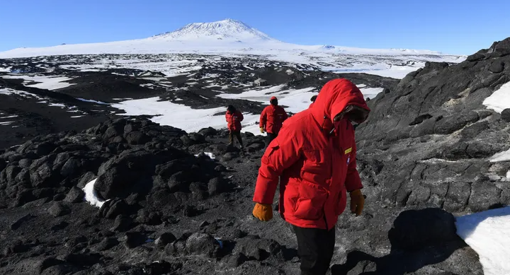
بركان ينفت الذهب في أقصى جنوب الأرض.. ما القصة؟
|
|
|
|
|
|
شعبة الخزانة والمتحف العلوي تشارك بثلاثة بحوث علمية في مؤتمر التراث الدولي الأول
|
|
|
|
لتطوير المهارات الهندسية والفنية للخدم .. العتبة العلوية المقدسة توفد كوادرها المتخصصين لورش شركة (تويوتا)
|
|
|
|
برعاية أبوية من العتبة العلوية المقدسة .. مشروع قنبر (2) السكني يدخل حيز التنفيذ
|
|
|
|
العتبة العلوية المقدسة تقيم برنامجاً حافلاً لنخبة من طلبة ثانوية المتفوقين الثانية في النجف الأشرف
|